WB
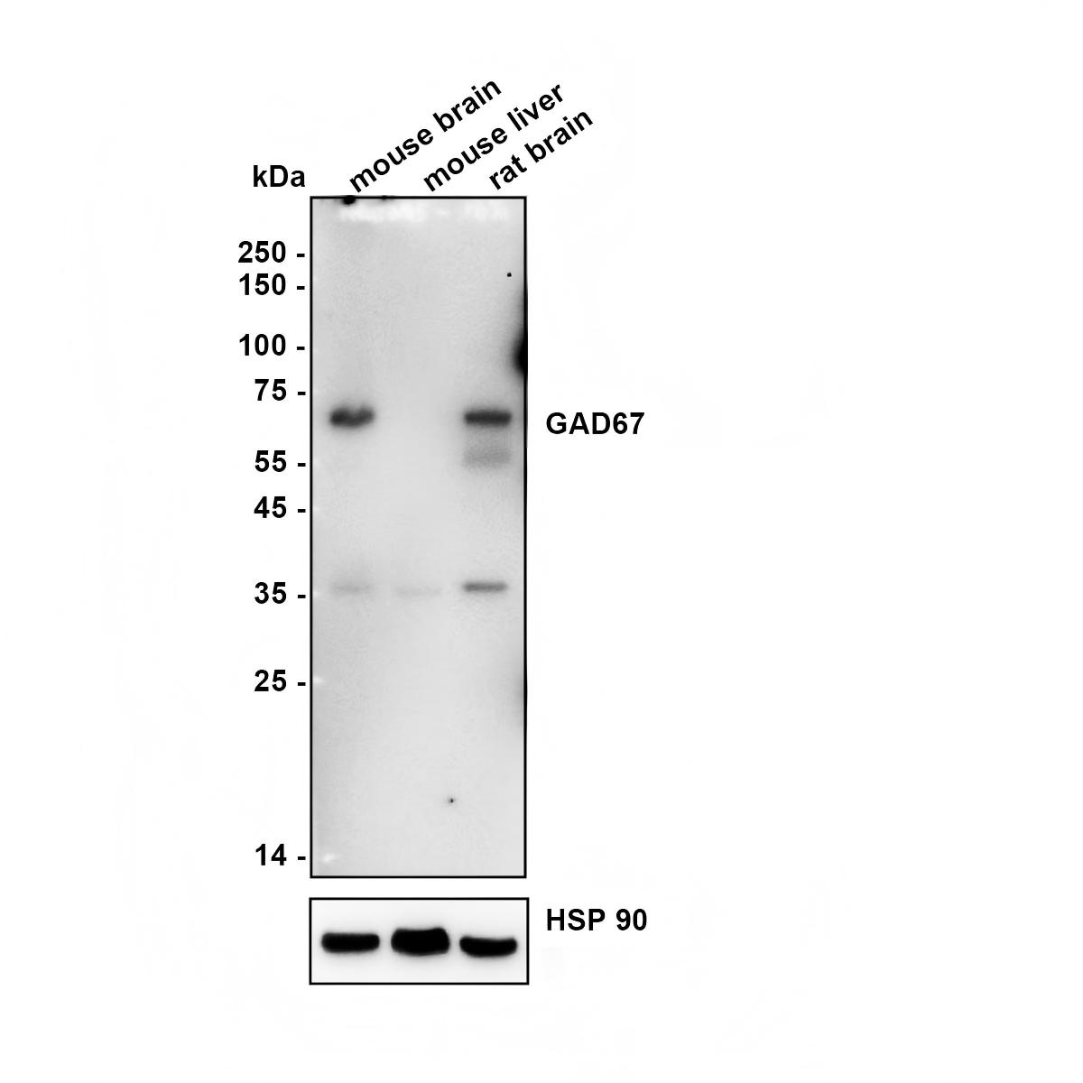
Western blot analysis of GAD67 on different lysates with Rabbit anti-GAD67 antibody at 1/5,000 dilution.Lysates/proteins at 20 µg/Lane.Exposure time: 3 minutes;Mouse liver is a negative control .Blocking: 5% NFDM/TBST, 1 hour at room temperature Primary antibody: HA724362, 1/5,000 in primary antibody dilution buffer , overnight at 4 °C Secondary antibody: Goat anti-Rabbit IgG-HRP, 1/50,000 in 5% NFDM/TBST, 1 hour at room temperatureIHC
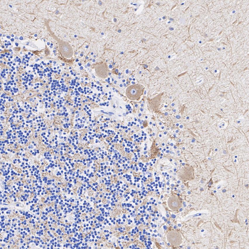
Application: Immunohistochemistry (IHC-P). Species: Human. Tissue: Cerebellum. Sample: Paraffin-embedded section. Antigen retrieval: Heat-mediated, Tris-EDTA buffer (pH 9.0), 20 minutes at 95°C. Wash buffer: 1× TBST. Endogenous peroxidase blocking: 3% H₂O₂, 10 minutes at room temperature. Blocking: 1% BSA + 10% normal goat serum, 10 minutes at room temperature. Primary antibody: 1/1,000, 1 hour at room temperature. Secondary antibody: 20 minutes at room temperature.IHC
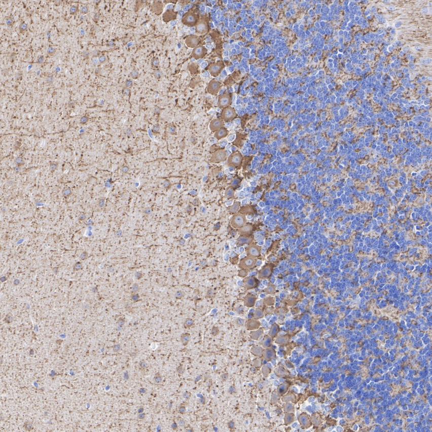
Application: Immunohistochemistry (IHC-P). Species: Mouse. Tissue: Cerebellum. Sample: Paraffin-embedded section. Antigen retrieval: Heat-mediated, Tris-EDTA buffer (pH 9.0), 20 minutes at 95°C. Wash buffer: 1× TBST. Endogenous peroxidase blocking: 3% H₂O₂, 10 minutes at room temperature. Blocking: 1% BSA + 10% normal goat serum, 10 minutes at room temperature. Primary antibody: 1/1,000, 1 hour at room temperature. Secondary antibody: 20 minutes at room temperature.IHC
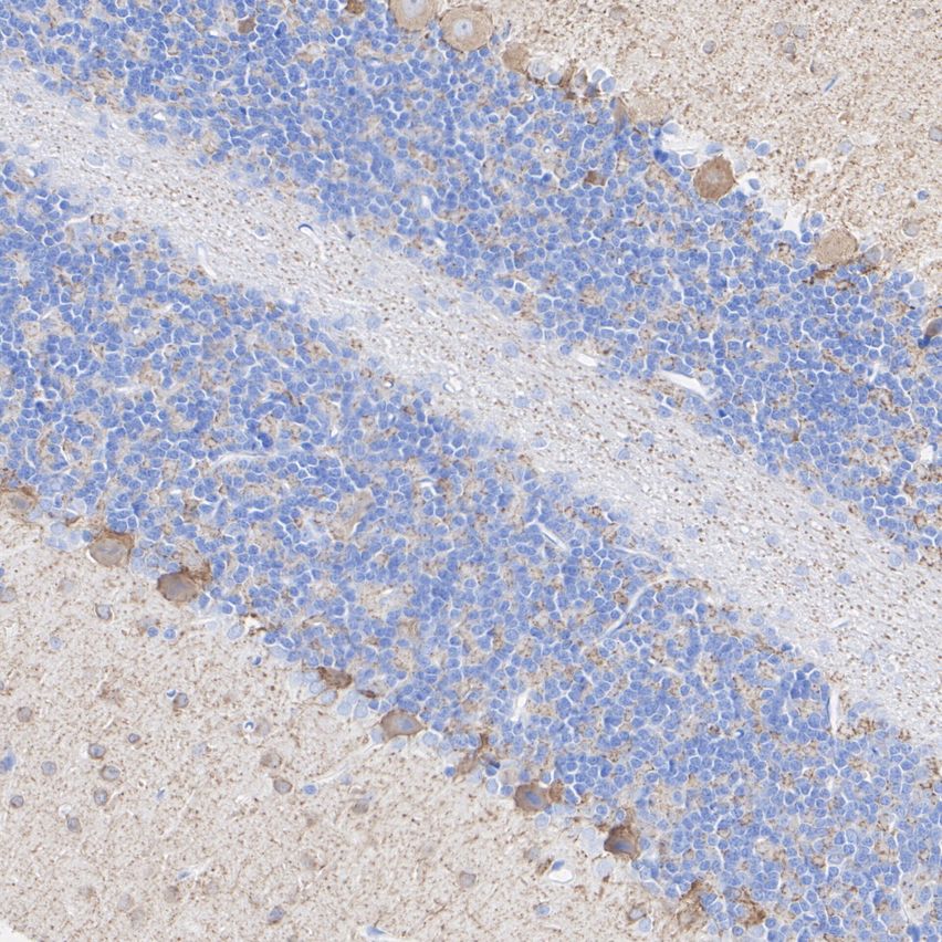
Application: Immunohistochemistry (IHC-P). Species: Rat. Tissue: Cerebellum. Sample: Paraffin-embedded section. Antigen retrieval: Heat-mediated, Tris-EDTA buffer (pH 9.0), 20 minutes at 95°C. Wash buffer: 1× TBST. Endogenous peroxidase blocking: 3% H₂O₂, 10 minutes at room temperature. Blocking: 1% BSA + 10% normal goat serum, 10 minutes at room temperature. Primary antibody: 1/1,000, 1 hour at room temperature. Secondary antibody: 20 minutes at room temperature.IF-P
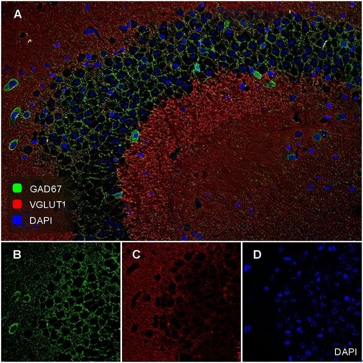
Application: Immunofluorescence (IF-tissue). Species: Mouse. Tissue: Hippocampus. Sample: Paraffin-embedded section. Antigen retrieval: Heat-mediated, Tris-EDTA buffer (pH 9.0), 20 minutes at 95°C. Wash buffer: 1× PBST. Endogenous peroxidase blocking: 3% H2O2, 10 minutes. Blocking: 1% BSA + 10% normal goat serum, 10 minutes at room temperature. Primary antibody: GAD67, 1/500 (Rabbit, Green), VGLUT1, 1/500 (Guinea pig, Red), overnight at 4℃. Secondary antibody: Goat Anti-Rabbit IgG (488), Goat anti-Guinea pig IgG (594), 1.5 hours at room temperature.IF-P
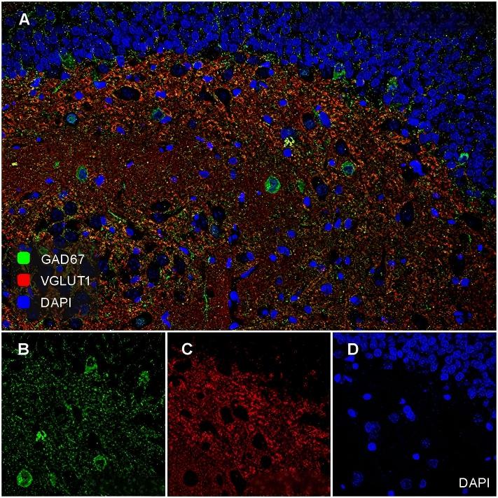
Application: Immunofluorescence (IF-tissue). Species: Rat. Tissue: Hippocampus. Sample: Paraffin-embedded section. Antigen retrieval: Heat-mediated, Tris-EDTA buffer (pH 9.0), 20 minutes at 95°C. Wash buffer: 1× PBST. Endogenous peroxidase blocking: 3% H2O2, 10 minutes. Blocking: 1% BSA + 10% normal goat serum, 10 minutes at room temperature. Primary antibody: GAD67, 1/500 (Rabbit, Green), VGLUT1, 1/500 (Guinea pig, Red), overnight at 4℃. Secondary antibody: Goat Anti-Rabbit IgG (488), Goat anti-Guinea pig IgG (594), 1.5 hours at room temperature.| Product Name | GAD67 Recombinant Antibody - Rabbit IgG (Chimeric) |
|---|---|
| Antibody Type | Primary Antibodies |
| Immunogen | Recombinant protein within human GAD67 aa 1-594. |
| Clonality | Monoclonal |
|---|---|
| Isotype | IgG |
| Tested Applications | IF-PIHCWB |
| WB:1:5000 IHC:1:1000 IF-P:1:500 |
|
| Species Reactivity | HumanMouseRat |
| Concentration | 1mg/ml |
| Purification | Protein A |
| Gene Symbol | GAD1 |
|---|---|
| Gene Synonyms | GAD SCP CPSQ1 DEE89 GAD-67 |
| Gene Full Name | glutamate decarboxylase 1 |
| Gene Summary | This gene encodes one of several forms of glutamic acid decarboxylase, identified as a major autoantigen in insulin-dependent diabetes. The enzyme encoded is responsible for catalyzing the production of gamma-aminobutyric acid from L-glutamic acid. A pathogenic role for this enzyme has been identified in the human pancreas since it has been identified as an autoantigen and an autoreactive T cell target in insulin-dependent diabetes. This gene may also play a role in the stiff man syndrome. Deficiency in this enzyme has been shown to lead to pyridoxine dependency with seizures. Alternative splicing of this gene results in two products, the predominant 67-kD form and a less-frequent 25-kD form. [provided by RefSeq, Jul 2008] |
| Alternative Names | 67 kDa glutamic acid decarboxylase antibody CPSQ1 antibody DCE1 antibody DCE1_HUMAN antibody EC 4.1.1.15 antibody FLJ45882 antibody GAD 67 antibody GAD antibody GAD-67 antibody GAD1 antibody 67 kDa glutamic acid decarboxylase antibody CPSQ1 antibody DCE1 antibody DCE1_HUMAN antibody EC 4.1.1.15 antibody FLJ45882 antibody GAD 67 antibody GAD antibody GAD-67 antibody GAD1 antibody Glutamate decarboxylase 1 (brain 67kDa) antibody Glutamate decarboxylase 1 antibody Glutamate decarboxylase 1 brain 67kD antibody Glutamate decarboxylase 1 brain 67kDa antibody Glutamate decarboxylase 67 kDa isoform antibody Glutamate decarboxylase brain 67-KD antibody OTTHUMP00000041055 antibody SCP antibody |
| Molecular Weight(MW) | 67 kDa |
| Cellular Localization | Cytoplasm, Plasma Membrane. |
WB
Western blot analysis of GAD67 on different lysates with Rabbit anti-GAD67 antibody at 1/5,000 dilution.Lysates/proteins at 20 µg/Lane.Exposure time: 3 minutes;Mouse liver is a negative control .Blocking: 5% NFDM/TBST, 1 hour at room temperature Primary antibody: HA724362, 1/5,000 in primary antibody dilution buffer , overnight at 4 °C Secondary antibody: Goat anti-Rabbit IgG-HRP, 1/50,000 in 5% NFDM/TBST, 1 hour at room temperature
IHC
Application: Immunohistochemistry (IHC-P). Species: Human. Tissue: Cerebellum. Sample: Paraffin-embedded section. Antigen retrieval: Heat-mediated, Tris-EDTA buffer (pH 9.0), 20 minutes at 95°C. Wash buffer: 1× TBST. Endogenous peroxidase blocking: 3% H₂O₂, 10 minutes at room temperature. Blocking: 1% BSA + 10% normal goat serum, 10 minutes at room temperature. Primary antibody: 1/1,000, 1 hour at room temperature. Secondary antibody: 20 minutes at room temperature.
IHC
Application: Immunohistochemistry (IHC-P). Species: Mouse. Tissue: Cerebellum. Sample: Paraffin-embedded section. Antigen retrieval: Heat-mediated, Tris-EDTA buffer (pH 9.0), 20 minutes at 95°C. Wash buffer: 1× TBST. Endogenous peroxidase blocking: 3% H₂O₂, 10 minutes at room temperature. Blocking: 1% BSA + 10% normal goat serum, 10 minutes at room temperature. Primary antibody: 1/1,000, 1 hour at room temperature. Secondary antibody: 20 minutes at room temperature.
IHC
Application: Immunohistochemistry (IHC-P). Species: Rat. Tissue: Cerebellum. Sample: Paraffin-embedded section. Antigen retrieval: Heat-mediated, Tris-EDTA buffer (pH 9.0), 20 minutes at 95°C. Wash buffer: 1× TBST. Endogenous peroxidase blocking: 3% H₂O₂, 10 minutes at room temperature. Blocking: 1% BSA + 10% normal goat serum, 10 minutes at room temperature. Primary antibody: 1/1,000, 1 hour at room temperature. Secondary antibody: 20 minutes at room temperature.
IF-P
Application: Immunofluorescence (IF-tissue). Species: Mouse. Tissue: Hippocampus. Sample: Paraffin-embedded section. Antigen retrieval: Heat-mediated, Tris-EDTA buffer (pH 9.0), 20 minutes at 95°C. Wash buffer: 1× PBST. Endogenous peroxidase blocking: 3% H2O2, 10 minutes. Blocking: 1% BSA + 10% normal goat serum, 10 minutes at room temperature. Primary antibody: GAD67, 1/500 (Rabbit, Green), VGLUT1, 1/500 (Guinea pig, Red), overnight at 4℃. Secondary antibody: Goat Anti-Rabbit IgG (488), Goat anti-Guinea pig IgG (594), 1.5 hours at room temperature.
IF-P
Application: Immunofluorescence (IF-tissue). Species: Rat. Tissue: Hippocampus. Sample: Paraffin-embedded section. Antigen retrieval: Heat-mediated, Tris-EDTA buffer (pH 9.0), 20 minutes at 95°C. Wash buffer: 1× PBST. Endogenous peroxidase blocking: 3% H2O2, 10 minutes. Blocking: 1% BSA + 10% normal goat serum, 10 minutes at room temperature. Primary antibody: GAD67, 1/500 (Rabbit, Green), VGLUT1, 1/500 (Guinea pig, Red), overnight at 4℃. Secondary antibody: Goat Anti-Rabbit IgG (488), Goat anti-Guinea pig IgG (594), 1.5 hours at room temperature.| Application Notes | WB:1:5000 IHC:1:1000 IF-P:1:500 |
|---|
| Form | Liquid |
|---|---|
| Storage Instructions | Shipped at 4℃. Store at +4℃ short term (1-2 weeks). It is recommended to aliquot into single-use upon delivery. Store at -20℃ long term. |
| Storage Buffer | PBS (pH7.4), 0.1% BSA, 40% Glycerol, 0.2% Proclean 950. |


