WB
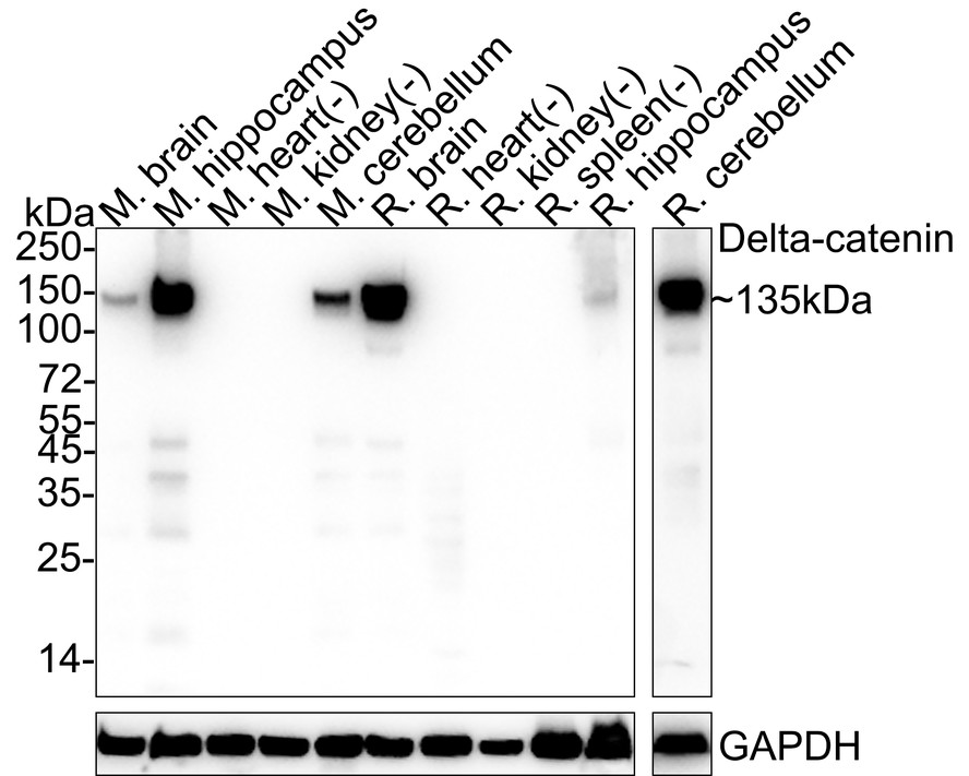
Western blot analysis of Delta-catenin on different lysates with Rabbit anti-Delta-catenin antibody at 1/1,000 dilution. Lysates/proteins at 40 µg/Lane. Exposure time: 1 minute 2 seconds; 4-20% SDS-PAGE gel. Proteins were transferred to a PVDF membrane and blocked with 5% NFDM/TBST for 1 hour at room temperature. The primary antibody at 1/1,000 dilution was used in 5% NFDM/TBST at 4℃ overnight. Goat Anti-Rabbit IgG - HRP Secondary Antibody at 1/50,000 dilution was used for 1 hour at room temperature.IP
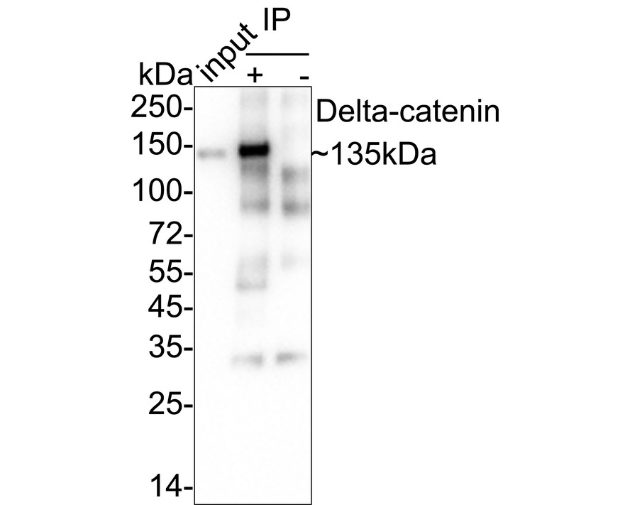
Delta-catenin was immunoprecipitated from 0.2 mg mouse brain tissue lysate with Rabbit anti-Delta-catenin antibody at 2 µg/10 µl beads. Western blot was performed from the immunoprecipitate using Rabbit anti-Delta-catenin antibody at 1/1,000 dilution. Anti-Rabbit IgG for IP Nano-secondary antibody at 1/5,000 dilution was used for 1 hour at room temperature. Lane 1: Mouse brain tissue lysate (input) Lane 2: Rabbit anti-Delta-catenin antibody IP in mouse brain tissue lysate Lane 3: Rabbit IgG instead of Rabbit anti-Delta-catenin antibody in mouse brain tissue lysate Blocking/Dilution buffer: 5% NFDM/TBST Exposure time: 17 seconds;IHC
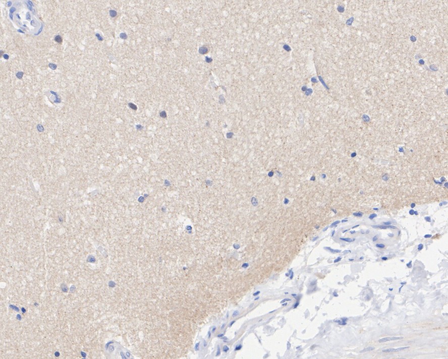
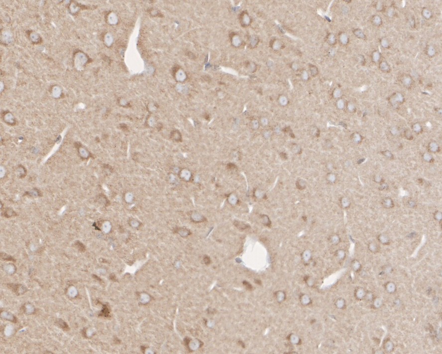
Immunohistochemical analysis of paraffin-embedded human brain tissue with Rabbit anti-Delta-catenin antibody at 1/200 dilution. The section was pre-treated using heat mediated antigen retrieval with sodium citrate buffer (pH 6.0) (high pressure) for 2 minutes. The tissues were blocked in 1% BSA for 20 minutes at room temperature, washed with ddH2O and PBS, and then probed with the primary antibody at 1/200 dilution for 1 hour at room temperature. The detection was performed using an HRP conjugated compact polymer system. DAB was used as the chromogen. Tissues were counterstained with hematoxylin and mounted with DPX.IHC
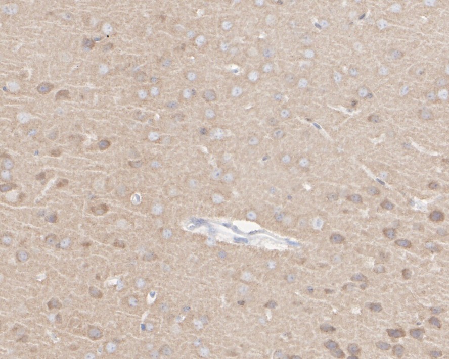
Immunohistochemical analysis of paraffin-embedded mouse brain tissue with Rabbit anti-Delta-catenin antibody at 1/200 dilution. The section was pre-treated using heat mediated antigen retrieval with sodium citrate buffer (pH 6.0) (high pressure) for 2 minutes. The tissues were blocked in 1% BSA for 20 minutes at room temperature, washed with ddH2O and PBS, and then probed with the primary antibody at 1/200 dilution for 1 hour at room temperature. The detection was performed using an HRP conjugated compact polymer system. DAB was used as the chromogen. Tissues were counterstained with hematoxylin and mounted with DPX.IHC
Immunohistochemical analysis of paraffin-embedded rat brain tissue with Rabbit anti-Delta-catenin antibody at 1/200 dilution. The section was pre-treated using heat mediated antigen retrieval with sodium citrate buffer (pH 6.0) (high pressure) for 2 minutes. The tissues were blocked in 1% BSA for 20 minutes at room temperature, washed with ddH2O and PBS, and then probed with the primary antibody at 1/200 dilution for 1 hour at room temperature. The detection was performed using an HRP conjugated compact polymer system. DAB was used as the chromogen. Tissues were counterstained with hematoxylin and mounted with DPX.IF-F
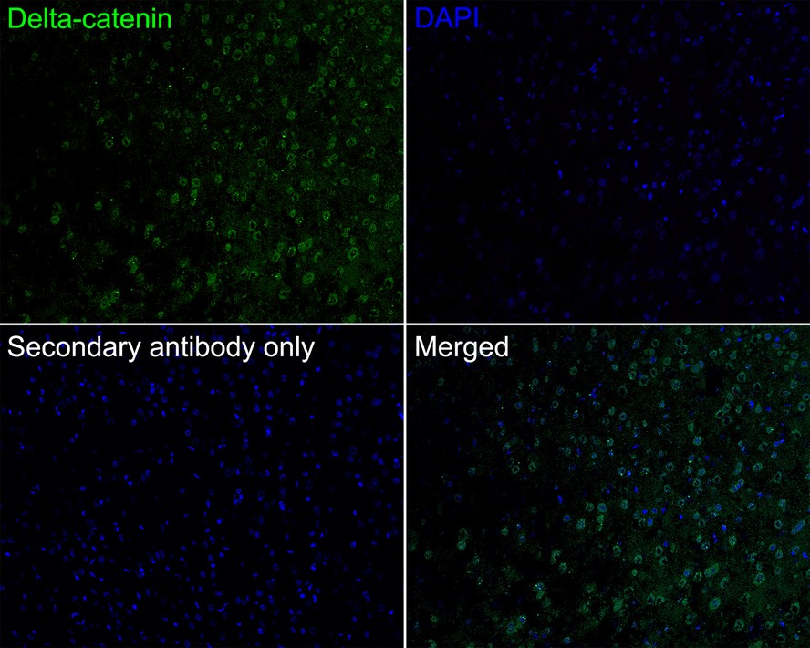
Application: IHC-Fr. Species: Rat. Site: Cerebral cortex. Sample: Frozen section. Antibody concentration: 1/200. Antigen retrieval: The section was pre-treated using heat mediated antigen retrieval with sodium citrate buffer (pH 6.0) for about 2 minutes in microwave oven.| Product Name | Delta-catenin Recombinant Rabbit Monoclonal Antibody |
|---|---|
| Antibody Type | Primary Antibodies |
| Immunogen | Recombinant protein within Mouse Catenin delta-2 aa 1-100 / 1,247. |
| Clonality | Monoclonal |
|---|---|
| Isotype | IgG |
| Host Species | Rabbit |
| Tested Applications | IF-FIHCIPWB |
| WB:1:1000 IHC:1:200 IP:1-2μg/sample IF-F:1:200 |
|
| Species Reactivity | HumanMouseRat |
| Concentration | 1mg/ml |
| Purification | Protein A |
| Gene Symbol | CTNND2 |
|---|---|
| Gene Synonyms | GT24 NPRAP |
| Gene Full Name | catenin delta 2 |
| Gene Summary | This gene encodes an adhesive junction associated protein of the armadillo/beta-catenin superfamily and is implicated in brain and eye development and cancer formation. The protein encoded by this gene promotes the disruption of E-cadherin based adherens junction to favor cell spreading upon stimulation by hepatocyte growth factor. This gene is overexpressed in prostate adenocarcinomas and is associated with decreased expression of tumor suppressor E-cadherin in this tissue. This gene resides in a region of the short arm of chromosome 5 that is deleted in Cri du Chat syndrome. Alternative splicing results in multiple transcript variants encoding different isoforms. [provided by RefSeq, Dec 2013] |
| Alternative Names | ARM-repeat protein antibody Catenin (cadherin associated protein) delta 2 antibody catenin (cadherin-associated protein) delta 2 (neural plakophilin-related arm-repeat protein) antibody Catenin delta 2 antibody Catenin delta-2 antibody CTND2_HUMAN antibody CTNND 2 antibody Ctnnd2 antibody Delta catenin antibody Delta-catenin antibody ARM-repeat protein antibody Catenin (cadherin associated protein) delta 2 antibody catenin (cadherin-associated protein) delta 2 (neural plakophilin-related arm-repeat protein) antibody Catenin delta 2 antibody Catenin delta-2 antibody CTND2_HUMAN antibody CTNND 2 antibody Ctnnd2 antibody Delta catenin antibody Delta-catenin antibody GT 24 antibody GT24 antibody Neural plakophilin related arm repeat protein antibody Neural plakophilin related armadillo repeat protein antibody Neural plakophilin-related ARM-repeat protein antibody Neural plakophilin-related Neurojungin antibody Neurojungin antibody NPRAP antibody T cell delta catenin antibody |
| Molecular Weight(MW) | 135 kDa |
| Cellular Localization | Nucleus, Cell junction, adherens junction, Cell projection, dendrite, Perikaryon. |
WB
Western blot analysis of Delta-catenin on different lysates with Rabbit anti-Delta-catenin antibody at 1/1,000 dilution. Lysates/proteins at 40 µg/Lane. Exposure time: 1 minute 2 seconds; 4-20% SDS-PAGE gel. Proteins were transferred to a PVDF membrane and blocked with 5% NFDM/TBST for 1 hour at room temperature. The primary antibody at 1/1,000 dilution was used in 5% NFDM/TBST at 4℃ overnight. Goat Anti-Rabbit IgG - HRP Secondary Antibody at 1/50,000 dilution was used for 1 hour at room temperature.
IP
Delta-catenin was immunoprecipitated from 0.2 mg mouse brain tissue lysate with Rabbit anti-Delta-catenin antibody at 2 µg/10 µl beads. Western blot was performed from the immunoprecipitate using Rabbit anti-Delta-catenin antibody at 1/1,000 dilution. Anti-Rabbit IgG for IP Nano-secondary antibody at 1/5,000 dilution was used for 1 hour at room temperature. Lane 1: Mouse brain tissue lysate (input) Lane 2: Rabbit anti-Delta-catenin antibody IP in mouse brain tissue lysate Lane 3: Rabbit IgG instead of Rabbit anti-Delta-catenin antibody in mouse brain tissue lysate Blocking/Dilution buffer: 5% NFDM/TBST Exposure time: 17 seconds;
IHC
Immunohistochemical analysis of paraffin-embedded human brain tissue with Rabbit anti-Delta-catenin antibody at 1/200 dilution. The section was pre-treated using heat mediated antigen retrieval with sodium citrate buffer (pH 6.0) (high pressure) for 2 minutes. The tissues were blocked in 1% BSA for 20 minutes at room temperature, washed with ddH2O and PBS, and then probed with the primary antibody at 1/200 dilution for 1 hour at room temperature. The detection was performed using an HRP conjugated compact polymer system. DAB was used as the chromogen. Tissues were counterstained with hematoxylin and mounted with DPX.
IHC
Immunohistochemical analysis of paraffin-embedded mouse brain tissue with Rabbit anti-Delta-catenin antibody at 1/200 dilution. The section was pre-treated using heat mediated antigen retrieval with sodium citrate buffer (pH 6.0) (high pressure) for 2 minutes. The tissues were blocked in 1% BSA for 20 minutes at room temperature, washed with ddH2O and PBS, and then probed with the primary antibody at 1/200 dilution for 1 hour at room temperature. The detection was performed using an HRP conjugated compact polymer system. DAB was used as the chromogen. Tissues were counterstained with hematoxylin and mounted with DPX.
IHC
Immunohistochemical analysis of paraffin-embedded rat brain tissue with Rabbit anti-Delta-catenin antibody at 1/200 dilution. The section was pre-treated using heat mediated antigen retrieval with sodium citrate buffer (pH 6.0) (high pressure) for 2 minutes. The tissues were blocked in 1% BSA for 20 minutes at room temperature, washed with ddH2O and PBS, and then probed with the primary antibody at 1/200 dilution for 1 hour at room temperature. The detection was performed using an HRP conjugated compact polymer system. DAB was used as the chromogen. Tissues were counterstained with hematoxylin and mounted with DPX.
IF-F
Application: IHC-Fr. Species: Rat. Site: Cerebral cortex. Sample: Frozen section. Antibody concentration: 1/200. Antigen retrieval: The section was pre-treated using heat mediated antigen retrieval with sodium citrate buffer (pH 6.0) for about 2 minutes in microwave oven.| Application Notes | WB:1:1000 IHC:1:200 IP:1-2μg/sample IF-F:1:200 |
|---|
| Form | Liquid |
|---|---|
| Storage Instructions | Shipped at 4℃. Store at +4℃ short term (1-2 weeks). It is recommended to aliquot into single-use upon delivery. Store at -20℃ long term. |
| Storage Buffer | TBS (pH7.4), 0.05% BSA, 40% Glycerol. Preservative: 0.05% Sodium Azide. |


