WB
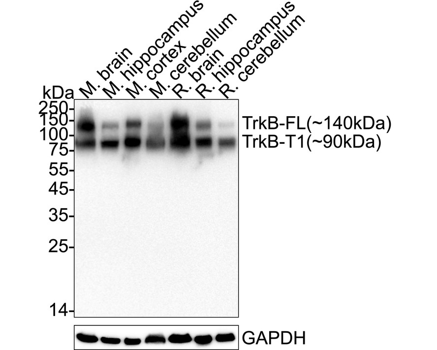
Western blot analysis of TrkB on different lysates with Rabbit anti-TrkB antibody at 1/5,000 dilution. Exposure time: 20 seconds; 4-20% SDS-PAGE gel. Proteins were transferred to a PVDF membrane and blocked with 5% NFDM/TBST for 1 hour at room temperature. The primary antibody at 1/5,000 dilution was used in 5% NFDM/TBST at 4℃ overnight. Goat Anti-Rabbit IgG - HRP Secondary Antibody at 1/50,000 dilution was used for 1 hour at room temperature.IHC
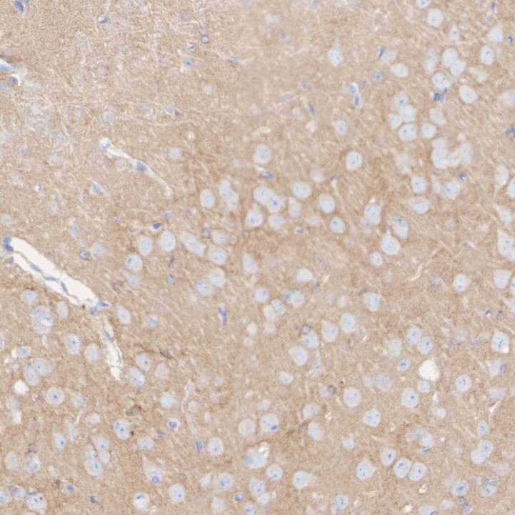
Immunohistochemical analysis of paraffin-embedded mouse brain tissue with Rabbit anti-TrkB antibody at 1/200 dilution. The section was pre-treated using heat mediated antigen retrieval with Tris-EDTA buffer (pH 9.0) for 20 minutes. The tissues were blocked in 1% BSA for 20 minutes at room temperature, washed with ddH2O and PBS, and then probed with the primary antibody at 1/200 dilution for 1 hour at room temperature. The detection was performed using an HRP conjugated compact polymer system. DAB was used as the chromogen. Tissues were counterstained with hematoxylin and mounted with DPX.IHC
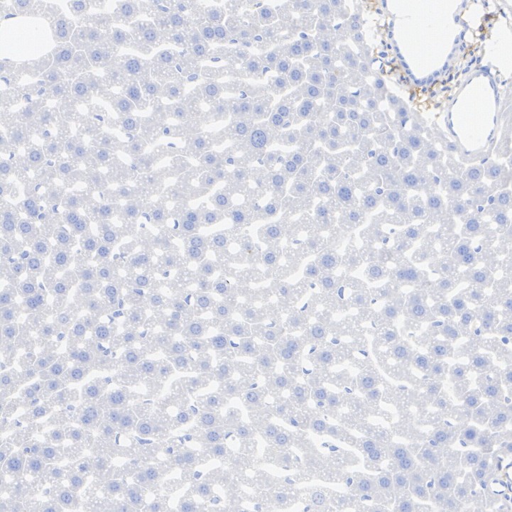
Immunohistochemical analysis of paraffin-embedded mouse liver tissue (negative) with Rabbit anti-TrkB antibody at 1/200 dilution. The section was pre-treated using heat mediated antigen retrieval with Tris-EDTA buffer (pH 9.0) for 20 minutes. The tissues were blocked in 1% BSA for 20 minutes at room temperature, washed with ddH2O and PBS, and then probed with the primary antibody at 1/200 dilution for 1 hour at room temperature. The detection was performed using an HRP conjugated compact polymer system. DAB was used as the chromogen. Tissues were counterstained with hematoxylin and mounted with DPX.IHC
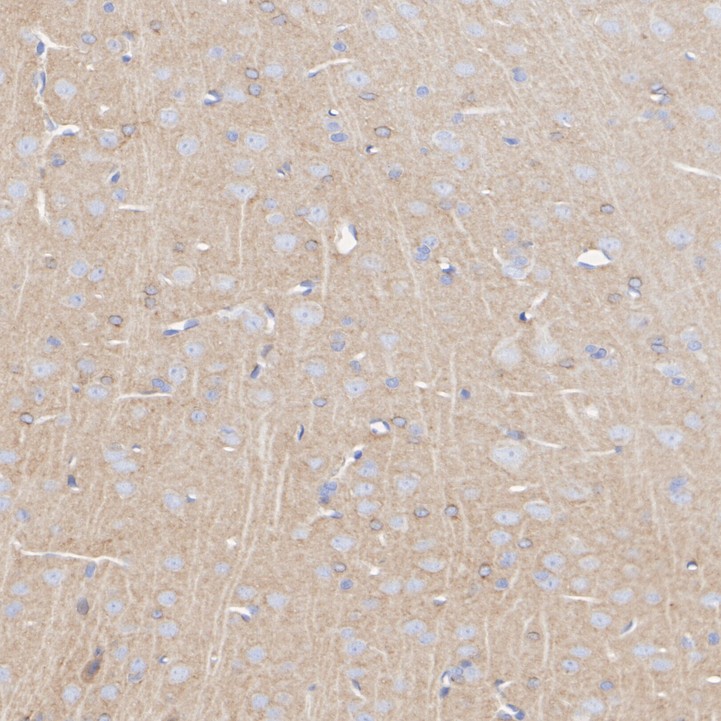
Immunohistochemical analysis of paraffin-embedded rat brain tissue with Rabbit anti-TrkB antibody at 1/200 dilution. The section was pre-treated using heat mediated antigen retrieval with Tris-EDTA buffer (pH 9.0) for 20 minutes. The tissues were blocked in 1% BSA for 20 minutes at room temperature, washed with ddH2O and PBS, and then probed with the primary antibody at 1/200 dilution for 1 hour at room temperature. The detection was performed using an HRP conjugated compact polymer system. DAB was used as the chromogen. Tissues were counterstained with hematoxylin and mounted with DPX.IF-F
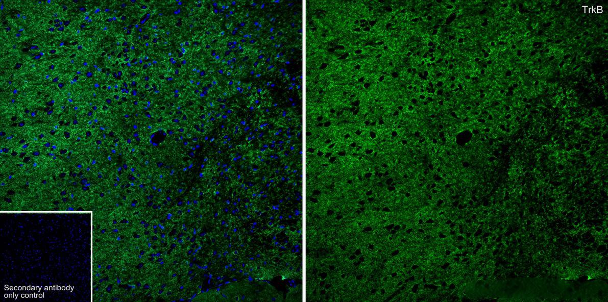
Application: Immunofluorescence (IHC-Fr). Species: Mouse. Tissue: Brain. Sample: Frozen section. Antigen retrieval: Not required. Wash buffer: 1× TBST. Blocking: 10% normal goat serum + 1% Triton X-100 + 0.3 M Glycine in TBST, 30 minutes at room temperature. Primary antibody:Rabbit anti-TrkB antibody,1/200, overnight at 4℃. Secondary antibody: Goat Anti-Rabbit IgG (488), 1.5 hours at room temperature.| Product Name | TrkB Recombinant Rabbit Monoclonal Antibody |
|---|---|
| Antibody Type | Primary Antibodies |
| Immunogen | Recombinant protein within human TrkB aa 100-450/822. |
| Clonality | Monoclonal |
|---|---|
| Isotype | IgG |
| Host Species | Rabbit |
| Tested Applications | IF-FIHCWB |
| WB:1:2000-1:5000 IHC:1:200 IF-F:1:200 |
|
| Species Reactivity | MouseRat |
| Concentration | 1mg/ml |
| Purification | Protein A |
| Gene Symbol | Ntrk2 |
|---|---|
| Gene Synonyms | Tkrb trkB trk-B GP145-TrkB/GP95-TrkB |
| Gene Full Name | neurotrophic tyrosine kinase, receptor, type 2 |
| Gene Summary | Enables brain-derived neurotrophic factor binding activity and brain-derived neurotrophic factor receptor activity. Involved in several processes, including brain-derived neurotrophic factor receptor signaling pathway; modulation of chemical synaptic transmission; and nervous system development. Acts upstream of or within several processes, including neurogenesis; neuronal action potential propagation; and positive regulation of peptidyl-serine phosphorylation. Located in several cellular components, including early endosome; postsynaptic density; and terminal bouton. Is expressed in several structures, including alimentary system; future brain; genitourinary system; integumental system; and nervous system. Human ortholog(s) of this gene implicated in several diseases, including alcohol dependence; autistic disorder; developmental and epileptic encephalopathy 58; heroin dependence; and morbid obesity. Orthologous to human NTRK2 (neurotrophic receptor tyrosine kinase 2). [provided by Alliance of Genome Resources, Jul 2025] |
| Alternative Names | AI848316 antibody BDNF tropomyosine receptor kinase B antibody BDNF/NT 3 growth factors receptor antibody BDNF/NT-3 growth factors receptor antibody Brain derived neurotrophic factor receptor antibody C030027L06Rik antibody EC 2.7.10.1 antibody GP145 TrkB antibody GP145-TrkB antibody GP145-TrkB/GP95-TrkB antibody AI848316 antibody BDNF tropomyosine receptor kinase B antibody BDNF/NT 3 growth factors receptor antibody BDNF/NT-3 growth factors receptor antibody Brain derived neurotrophic factor receptor antibody C030027L06Rik antibody EC 2.7.10.1 antibody GP145 TrkB antibody GP145-TrkB antibody GP145-TrkB/GP95-TrkB antibody GP95 TrkB antibody Neurotrophic receptor tyrosine kinase 2 antibody Neurotrophic tyrosine kinase receptor type 2 antibody Neurotrophin receptor tyrosine kinase type 2 antibody NTRK 2 antibody Ntrk2 antibody NTRK2_HUMAN antibody Obesity hyperphagia and developmental delay included antibody RATTRKB1 antibody Tkrb antibody Trk B antibody Trk-B antibody TRKB antibody TrkB tyrosine kinase antibody TRKB1 antibody Tropomyosin related kinase B antibody tyrosine kinase receptor B antibody Tyrosine receptor kinase B antibody |
| Molecular Weight(MW) | 92 kDa(Observed band size: 140/90 kDa) |
| Cellular Localization | Cell membrane, Endosome membrane, Early endosome membrane, axon, dendrite, perinuclear region, postsynaptic density. |
WB
Western blot analysis of TrkB on different lysates with Rabbit anti-TrkB antibody at 1/5,000 dilution. Exposure time: 20 seconds; 4-20% SDS-PAGE gel. Proteins were transferred to a PVDF membrane and blocked with 5% NFDM/TBST for 1 hour at room temperature. The primary antibody at 1/5,000 dilution was used in 5% NFDM/TBST at 4℃ overnight. Goat Anti-Rabbit IgG - HRP Secondary Antibody at 1/50,000 dilution was used for 1 hour at room temperature.
IHC
Immunohistochemical analysis of paraffin-embedded mouse brain tissue with Rabbit anti-TrkB antibody at 1/200 dilution. The section was pre-treated using heat mediated antigen retrieval with Tris-EDTA buffer (pH 9.0) for 20 minutes. The tissues were blocked in 1% BSA for 20 minutes at room temperature, washed with ddH2O and PBS, and then probed with the primary antibody at 1/200 dilution for 1 hour at room temperature. The detection was performed using an HRP conjugated compact polymer system. DAB was used as the chromogen. Tissues were counterstained with hematoxylin and mounted with DPX.
IHC
Immunohistochemical analysis of paraffin-embedded mouse liver tissue (negative) with Rabbit anti-TrkB antibody at 1/200 dilution. The section was pre-treated using heat mediated antigen retrieval with Tris-EDTA buffer (pH 9.0) for 20 minutes. The tissues were blocked in 1% BSA for 20 minutes at room temperature, washed with ddH2O and PBS, and then probed with the primary antibody at 1/200 dilution for 1 hour at room temperature. The detection was performed using an HRP conjugated compact polymer system. DAB was used as the chromogen. Tissues were counterstained with hematoxylin and mounted with DPX.
IHC
Immunohistochemical analysis of paraffin-embedded rat brain tissue with Rabbit anti-TrkB antibody at 1/200 dilution. The section was pre-treated using heat mediated antigen retrieval with Tris-EDTA buffer (pH 9.0) for 20 minutes. The tissues were blocked in 1% BSA for 20 minutes at room temperature, washed with ddH2O and PBS, and then probed with the primary antibody at 1/200 dilution for 1 hour at room temperature. The detection was performed using an HRP conjugated compact polymer system. DAB was used as the chromogen. Tissues were counterstained with hematoxylin and mounted with DPX.
IF-F
Application: Immunofluorescence (IHC-Fr). Species: Mouse. Tissue: Brain. Sample: Frozen section. Antigen retrieval: Not required. Wash buffer: 1× TBST. Blocking: 10% normal goat serum + 1% Triton X-100 + 0.3 M Glycine in TBST, 30 minutes at room temperature. Primary antibody:Rabbit anti-TrkB antibody,1/200, overnight at 4℃. Secondary antibody: Goat Anti-Rabbit IgG (488), 1.5 hours at room temperature.| Application Notes | WB:1:2000-1:5000 IHC:1:200 IF-F:1:200 |
|---|
| Form | Liquid |
|---|---|
| Storage Instructions | Shipped at 4℃. Store at +4℃ short term (1-2 weeks). It is recommended to aliquot into single-use upon delivery. Store at -20℃ long term. |
| Storage Buffer | TBS (pH7.4), 0.05% BSA, 40% Glycerol. Preservative: 0.05% Sodium Azide. |


