WB
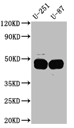
Western Blot: Positive WB detected in: U-251 whole cell lysate, U-87 whole cell lysate. All lanes: PAI1 Antibody at 1:1000. Secondary: Goat polyclonal to rabbit IgG at 1/50000 dilution.IHC
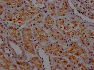
IHC image of SERPINE1 Recombinant Monoclonal Antibody diluted at 1:100 and staining in paraffin-embedded human kidney tissue performed on a Leica BondTM system. After dewaxing and hydration, antigen retrieval was mediated by high pressure in a citrate buffer (pH 6.0). Section was blocked with 10% normal goat serum 30min at RT. Then primary antibody (1% BSA) was incubated at 4℃ overnight. The primary is detected by a Goat anti-rabbit IgG polymer labeled by HRP and visualized using 0.05% DAB.| Product Name | SERPINE1 Recombinant Monoclonal Antibody |
|---|---|
| Antibody Type | Primary Antibodies |
| Immunogen | A synthesized peptide derived from human PAI1 |
| Clonality | Monoclonal |
|---|---|
| Isotype | IgG |
| Host Species | Rabbit |
| Tested Applications | IHCWB |
| WB:1:500-1:5000 IHC:1:50-1:200 |
|
| Species Reactivity | Human |
| Concentration | 1mg/ml |
| Purification | Affinity purified |
| Gene Symbol | SERPINE1 |
|---|---|
| Gene Synonyms | PAI PAI1 PAI-1 PLANH1 |
| Gene Full Name | serpin family E member 1 |
| Gene Summary | This gene encodes a member of the serine proteinase inhibitor (serpin) superfamily. This member is the principal inhibitor of tissue plasminogen activator (tPA) and urokinase (uPA), and hence is an inhibitor of fibrinolysis. The protein also functions as a component of innate antiviral immunity. Defects in this gene are the cause of plasminogen activator inhibitor-1 deficiency (PAI-1 deficiency), and high concentrations of the gene product are associated with thrombophilia. [provided by RefSeq, Aug 2020] |
| Alternative Names | Plasminogen activator inhibitor 1 (PAI) (PAI-1) (Endothelial plasminogen activator inhibitor) (Serpin E1) SERPINE1 PAI1 PLANH1 |
| Molecular Weight(MW) | 46, 44 kDa(Observed band size: 45 kDa) |
| Cellular Localization | Secreted. |
WB
Western Blot: Positive WB detected in: U-251 whole cell lysate, U-87 whole cell lysate. All lanes: PAI1 Antibody at 1:1000. Secondary: Goat polyclonal to rabbit IgG at 1/50000 dilution.
IHC
IHC image of SERPINE1 Recombinant Monoclonal Antibody diluted at 1:100 and staining in paraffin-embedded human kidney tissue performed on a Leica BondTM system. After dewaxing and hydration, antigen retrieval was mediated by high pressure in a citrate buffer (pH 6.0). Section was blocked with 10% normal goat serum 30min at RT. Then primary antibody (1% BSA) was incubated at 4℃ overnight. The primary is detected by a Goat anti-rabbit IgG polymer labeled by HRP and visualized using 0.05% DAB.| Application Notes | WB:1:500-1:5000 IHC:1:50-1:200 |
|---|
| Form | Liquid |
|---|---|
| Storage Instructions | Upon receipt, store at -20°C or -80°C. Avoid repeated freeze. |
| Storage Buffer | Rabbit IgG in 10mM phosphate buffered saline , pH 7.4, 150mM sodium chloride, 0.05% BSA, 0.02% sodium azide and 50% glycerol. |


