WB
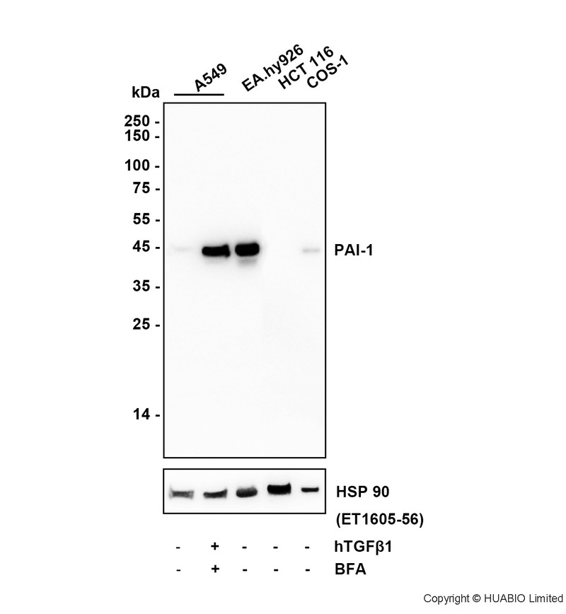
Western blot analysis of PAI1 on different lysates with Rabbit anti-PAI1 antibody at 1/5,000 dilution. Lane 1: A549 (Human lung adenocarcinoma cells) cell lysate Lane 2: A549 treated with 5ng/ml hTGFβ1 for 48 hours and 300ng/ml BFA for 16 hours cell lysate Lane 3: EA.hy926 (Human umbilical vein fusion cells) cell lysate Lane 4: HCT 116 (Human colon cancer cells) cell lysate Lane 5: COS-1 (African green monkey kidney fibroblasts) cell lysate Lysates/proteins at 15 µg/Lane. Exposure time: 30 seconds; ECL: K1801 HCT 116 is a negative control. Blocking: 5% NFDM/TBST, 1 hour at room temperature Primary antibody: 1/5,000 in primary antibody dilution buffer, overnight at 4 °C Secondary antibody: Goat anti-Rabbit IgG-HRP , 1/50,000 in 5% NFDM/TBST, 1 hour at room temperatureIP
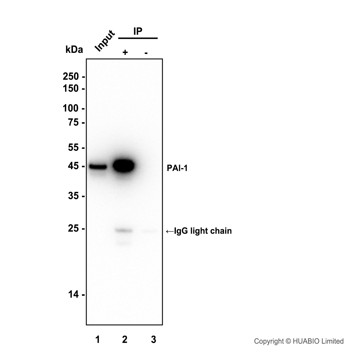
PAI1 was immunoprecipitated in 0.2 mg EA.hy926 (Human umbilical vein fusion cell) cell lysate with 2 µg/10 µl beads. Western blot was performed from the immunoprecipitate using PAI1 at 1/5,000 dilution. Anti-Rabbit IgG for IP Nano-secondary antibody at 1/5,000 dilution was used for 1 hour at room temperature. Lane 1: EA.hy926 cell lysate (input) Lane 2: IP in EA.hy926 cell lysate Lane 3: Rabbit IgG instead of HA724440 in EA.hy926 cell lysate Exposure time: 5 seconds Blocking: 5% NFDM/TBST, 1 hour at room temperature Primary dilution: 1/5,000 in primary antibody dilution buffer, 2 hours at room temperature| Product Name | PAI1 Recombinant Rabbit Monoclonal Antibody |
|---|---|
| Antibody Type | Primary Antibodies |
| Immunogen | Recombinant protein within human PAI1 aa 1-402. |
| Clonality | Monoclonal |
|---|---|
| Isotype | IgG |
| Host Species | Rabbit |
| Tested Applications | WB |
| WB:1:5000 IP:1-2μg/sample |
|
| Species Reactivity | HumanMonkeyMouse |
| Concentration | 1mg/ml |
| Purification | Protein A |
| Gene Symbol | SERPINE1 |
|---|---|
| Gene Synonyms | PAI PAI1 PAI-1 PLANH1 |
| Gene Full Name | serpin family E member 1 |
| Gene Summary | This gene encodes a member of the serine proteinase inhibitor (serpin) superfamily. This member is the principal inhibitor of tissue plasminogen activator (tPA) and urokinase (uPA), and hence is an inhibitor of fibrinolysis. The protein also functions as a component of innate antiviral immunity. Defects in this gene are the cause of plasminogen activator inhibitor-1 deficiency (PAI-1 deficiency), and high concentrations of the gene product are associated with thrombophilia. [provided by RefSeq, Aug 2020] |
| Alternative Names | Clade E antibody Endothelial plasminogen activator inhibitor antibody Nexin antibody Nexin plasminogen activator inhibitor type 1 antibody PAI 1 antibody PAI antibody PAI-1 antibody PAI1_HUMAN antibody PLANH1 antibody Plasminogen activator inhibitor 1 antibody Clade E antibody Endothelial plasminogen activator inhibitor antibody Nexin antibody Nexin plasminogen activator inhibitor type 1 antibody PAI 1 antibody PAI antibody PAI-1 antibody PAI1_HUMAN antibody PLANH1 antibody Plasminogen activator inhibitor 1 antibody Plasminogen activator inhibitor type 1 antibody Serine (or cysteine) proteinase inhibitor antibody Serine (or cysteine) proteinase inhibitor clade E (nexin plasminogen activator inhibitor type 1) member 1 antibody Serine proteinase inhibitor clade E member 1 antibody serpin antibody Serpin E1 antibody Serpin peptidase inhibitor clade E (nexin plasminogen activator inhibitor type 1) member 1 antibody Serpin peptidase inhibitor clade E antibody Serpine 1 antibody SERPINE1 antibody |
| Molecular Weight(MW) | 45 kDa(Observed band size: 45 kDa) |
| Cellular Localization | Secreted. |
WB
Western blot analysis of PAI1 on different lysates with Rabbit anti-PAI1 antibody at 1/5,000 dilution. Lane 1: A549 (Human lung adenocarcinoma cells) cell lysate Lane 2: A549 treated with 5ng/ml hTGFβ1 for 48 hours and 300ng/ml BFA for 16 hours cell lysate Lane 3: EA.hy926 (Human umbilical vein fusion cells) cell lysate Lane 4: HCT 116 (Human colon cancer cells) cell lysate Lane 5: COS-1 (African green monkey kidney fibroblasts) cell lysate Lysates/proteins at 15 µg/Lane. Exposure time: 30 seconds; ECL: K1801 HCT 116 is a negative control. Blocking: 5% NFDM/TBST, 1 hour at room temperature Primary antibody: 1/5,000 in primary antibody dilution buffer, overnight at 4 °C Secondary antibody: Goat anti-Rabbit IgG-HRP , 1/50,000 in 5% NFDM/TBST, 1 hour at room temperature
IP
PAI1 was immunoprecipitated in 0.2 mg EA.hy926 (Human umbilical vein fusion cell) cell lysate with 2 µg/10 µl beads. Western blot was performed from the immunoprecipitate using PAI1 at 1/5,000 dilution. Anti-Rabbit IgG for IP Nano-secondary antibody at 1/5,000 dilution was used for 1 hour at room temperature. Lane 1: EA.hy926 cell lysate (input) Lane 2: IP in EA.hy926 cell lysate Lane 3: Rabbit IgG instead of HA724440 in EA.hy926 cell lysate Exposure time: 5 seconds Blocking: 5% NFDM/TBST, 1 hour at room temperature Primary dilution: 1/5,000 in primary antibody dilution buffer, 2 hours at room temperature| Application Notes | WB:1:5000 IP:1-2μg/sample |
|---|
| Form | Liquid |
|---|---|
| Storage Instructions | Shipped at 4℃. Store at +4℃ short term (1-2 weeks). It is recommended to aliquot into single-use upon delivery. Store at -20℃ long term. |
| Storage Buffer | TBS (pH7.4), 0.05% BSA, 40% Glycerol. Preservative: 0.05% Sodium Azide. |


