WB
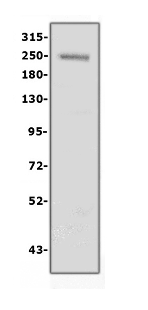
Western blot analysis of COL4A1 using anti-COL4A1 antibody. The sample well of each lane was loaded with 30 ug of sample under reducing conditions. Lane 1: human placenta tissue lysates. After electrophoresis, proteins were transferred to a membrane. Then the membrane was incubated with rabbit anti-COL4A1 antigen affinity purified polyclonal antibody at a dilution of 1:1000 and probed with a goat anti-rabbit IgG-HRP secondary antibody. The signal is developed using ECL Plus Western Blotting Substrate.IHC
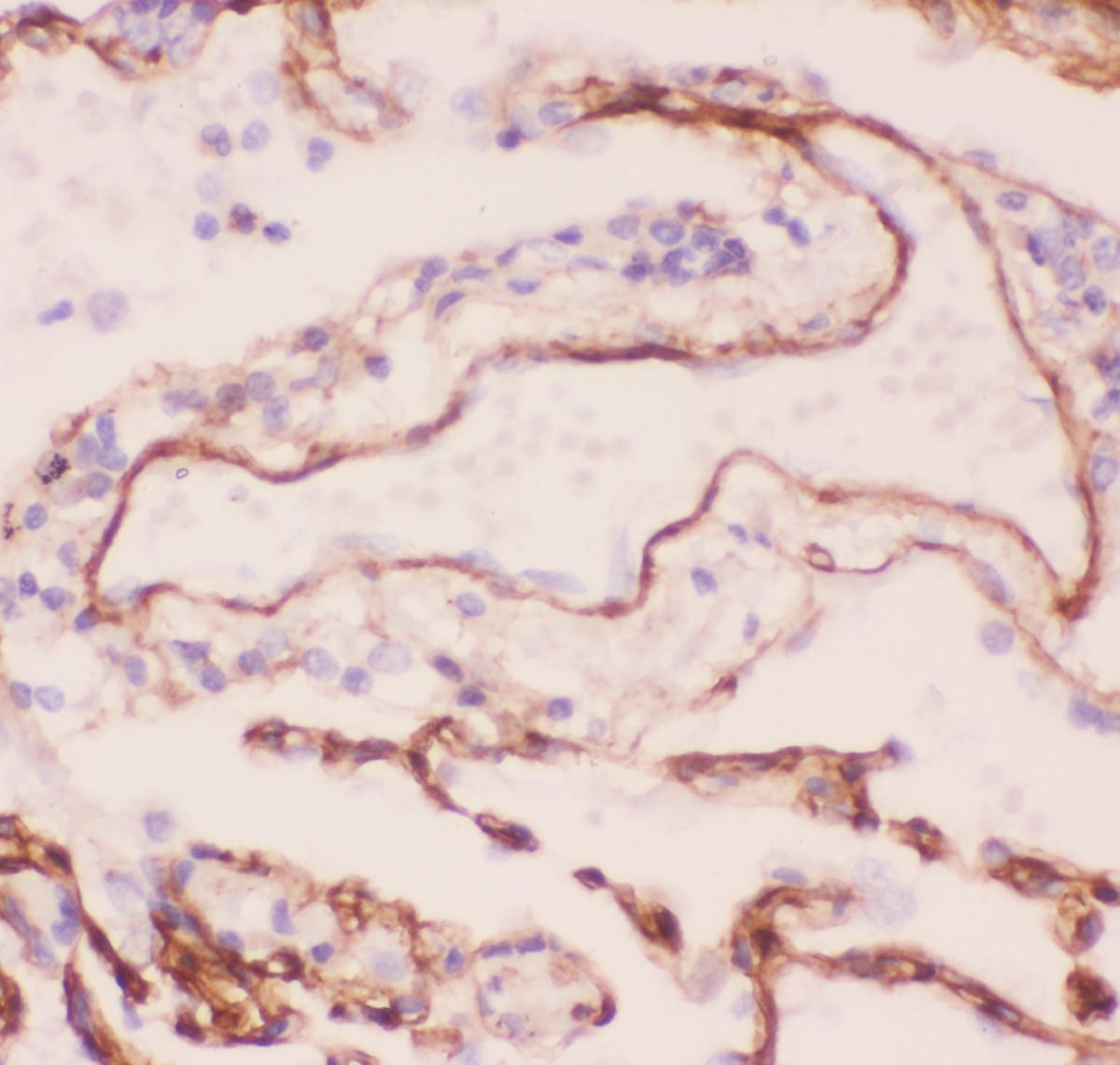
COL4A1 was detected in a paraffin-embedded section of human lung cancer tissue. Biotinylated goat anti-rabbit IgG was used as secondary antibody. The tissue section was incubated with rabbit anti-COL4A1 Antibody at a dilution of 1:200 and developed using Strepavidin-Biotin-Complex (SABC)with DAB as the chromogen.IF-P
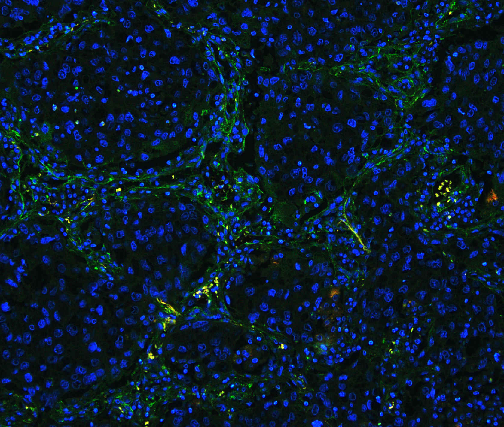
COL4A1 was detected in paraffin-embedded section of human lung cancer tissues. Heat mediated antigen retrieval was performed in citrate buffer (pH6, epitope retrieval solution ) for 20 mins. The tissue section was blocked with 10% goat serum. The tissue section was then incubated with 1μg/mL rabbit anti- COL4A1 Antibody overnight at 4°C. Biotin conjugated goat anti-rabbit IgG was used as secondary antibody and incubated for 30 minutes at 37°C. The tissue section was developed using Fluoro488 Conjugated Avidin. The section was counterstained with DAPI. Visualize using a fluorescence microscope and filter sets appropriate for the label used.| Product Name | Rabbit polyclonal antibody to COL4A1 |
|---|---|
| Antibody Type | Primary Antibodies |
| Immunogen | E.coli-derived human Collagen IV recombinant protein (Position: G1445-T1669). Human Collagen IV shares 97% amino acid (aa) sequence identity with mouse Collagen IV. |
| Clonality | Polyclonal |
|---|---|
| Isotype | IgG |
| Host Species | Rabbit |
| Tested Applications | IF-PIHCWB |
| WB:1:500-1:2000 IHC:1:50-1:400 IF-P:1:50-1:400 |
|
| Species Reactivity | HumanMouseRat |
| Concentration | 0.5mg/ml |
| Purification | Affinity purified |
| Gene Symbol | COL4A1 |
|---|---|
| Gene Synonyms | BSVD BSVD1 RATOR PADMAL COL4A1s |
| Gene Full Name | collagen type IV alpha 1 chain |
| Gene Summary | This gene encodes a type IV collagen alpha protein. Type IV collagen proteins are integral components of basement membranes. This gene shares a bidirectional promoter with a paralogous gene on the opposite strand. The protein consists of an amino-terminal 7S domain, a triple-helix forming collagenous domain, and a carboxy-terminal non-collagenous domain. It functions as part of a heterotrimer and interacts with other extracellular matrix components such as perlecans, proteoglycans, and laminins. In addition, proteolytic cleavage of the non-collagenous carboxy-terminal domain results in a biologically active fragment known as arresten, which has anti-angiogenic and tumor suppressor properties. Mutations in this gene cause porencephaly, cerebrovascular disease, and renal and muscular defects. Alternative splicing results in multiple transcript variants. [provided by RefSeq, Dec 2014] |
| Alternative Names | collagen type IV alpha 1 chain BSVD BSVD1s PADMAL RATOR COL4A1 |
| Molecular Weight(MW) | 161 kDa(Observed band size: 220 kDa) |
| Cellular Localization | Basement membrane,Extracellular matrix,Secreted. |
WB
Western blot analysis of COL4A1 using anti-COL4A1 antibody. The sample well of each lane was loaded with 30 ug of sample under reducing conditions. Lane 1: human placenta tissue lysates. After electrophoresis, proteins were transferred to a membrane. Then the membrane was incubated with rabbit anti-COL4A1 antigen affinity purified polyclonal antibody at a dilution of 1:1000 and probed with a goat anti-rabbit IgG-HRP secondary antibody. The signal is developed using ECL Plus Western Blotting Substrate.
IHC
COL4A1 was detected in a paraffin-embedded section of human lung cancer tissue. Biotinylated goat anti-rabbit IgG was used as secondary antibody. The tissue section was incubated with rabbit anti-COL4A1 Antibody at a dilution of 1:200 and developed using Strepavidin-Biotin-Complex (SABC)with DAB as the chromogen.
IF-P
COL4A1 was detected in paraffin-embedded section of human lung cancer tissues. Heat mediated antigen retrieval was performed in citrate buffer (pH6, epitope retrieval solution ) for 20 mins. The tissue section was blocked with 10% goat serum. The tissue section was then incubated with 1μg/mL rabbit anti- COL4A1 Antibody overnight at 4°C. Biotin conjugated goat anti-rabbit IgG was used as secondary antibody and incubated for 30 minutes at 37°C. The tissue section was developed using Fluoro488 Conjugated Avidin. The section was counterstained with DAPI. Visualize using a fluorescence microscope and filter sets appropriate for the label used.| Application Notes | WB:1:500-1:2000 IHC:1:50-1:400 IF-P:1:50-1:400 |
|---|
| Form | Liquid |
|---|---|
| Storage Instructions | 12 months from date of receipt, -20℃ as supplied. 6 months 2 to 8℃ after reconstitution. Avoid repeated freezing and thawing. |
| Storage Buffer | 500ug/ml antibody with PBS, 0.02% NaN3, 1 mg/ml BSA and 50% glycerol. |


