WB
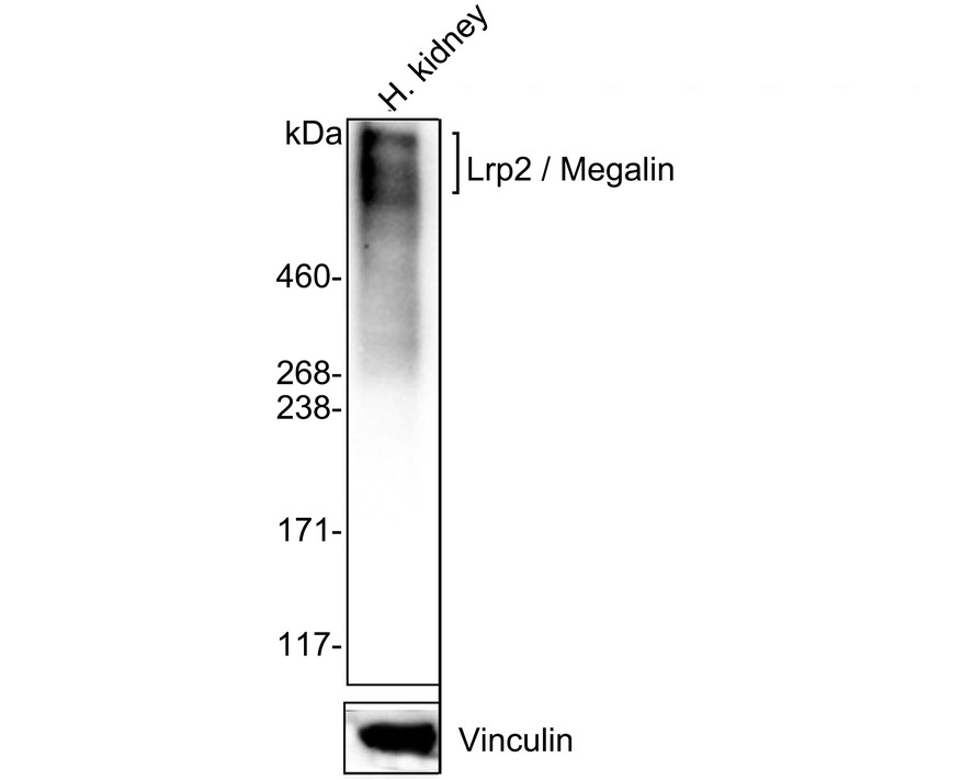
Western blot analysis of Lrp2 / Megalin on human kidney tissue lysates with Rabbit anti-Lrp2 / Megalin antibody at 1/2,000 dilution. Lysates/proteins at 40 µg/Lane. Exposure time: 2 minutes; 4-20% SDS-PAGE gel. Proteins were transferred to a PVDF membrane and blocked with 5% NFDM/TBST for 1 hour at room temperature. The primary antibody at 1/2,000 dilution was used in 5% NFDM/TBST at 4℃ overnight. Goat Anti-Rabbit IgG - HRP Secondary Antibody at 1/50,000 dilution was used for 1 hour at room temperature.IHC
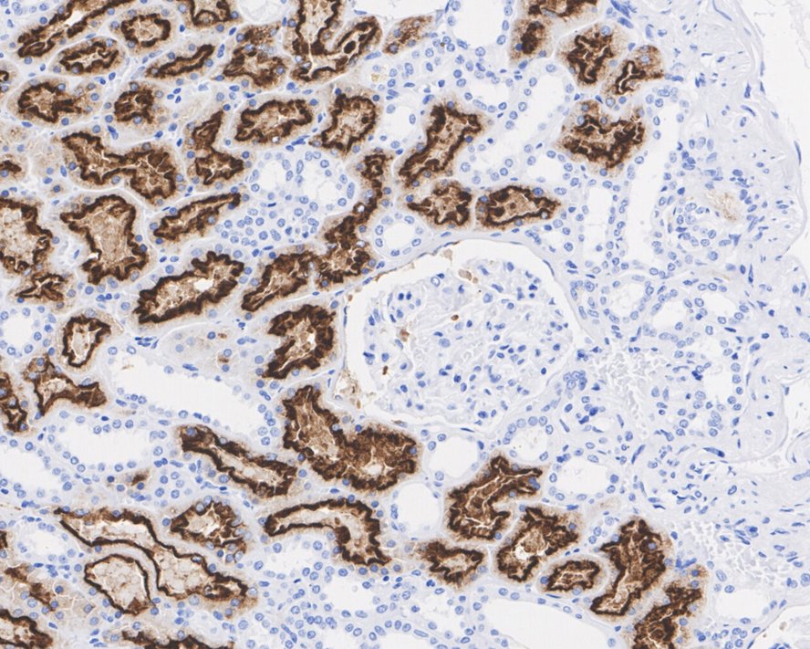
Immunohistochemical analysis of paraffin-embedded human kidney tissue with Rabbit anti-Lrp2 / Megalin antibody at 1/1000 dilution. The section was pre-treated using heat mediated antigen retrieval with Tris-EDTA buffer (pH 9.0) for 20 minutes. The tissues were blocked in 1% BSA for 20 minutes at room temperature, washed with ddH2O and PBS, and then probed with the primary antibody at 1/200 dilution for 1 hour at room temperature. The detection was performed using an HRP conjugated compact polymer system. DAB was used as the chromogen. Tissues were counterstained with hematoxylin and mounted with DPX.IHC
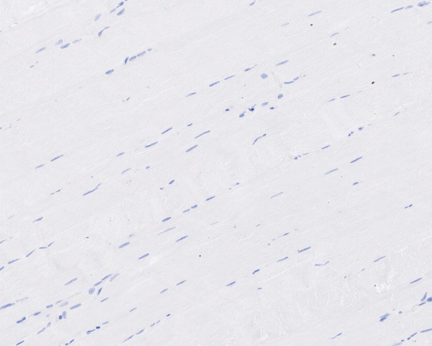
Immunohistochemical analysis of paraffin-embedded human skeletal muscle tissue (negative) with Rabbit anti-Lrp2 / Megalin antibody at 1/1000 dilution. The section was pre-treated using heat mediated antigen retrieval with Tris-EDTA buffer (pH 9.0) for 20 minutes. The tissues were blocked in 1% BSA for 20 minutes at room temperature, washed with ddH2O and PBS, and then probed with the primary antibody at 1/200 dilution for 1 hour at room temperature. The detection was performed using an HRP conjugated compact polymer system. DAB was used as the chromogen. Tissues were counterstained with hematoxylin and mounted with DPX.IF-P
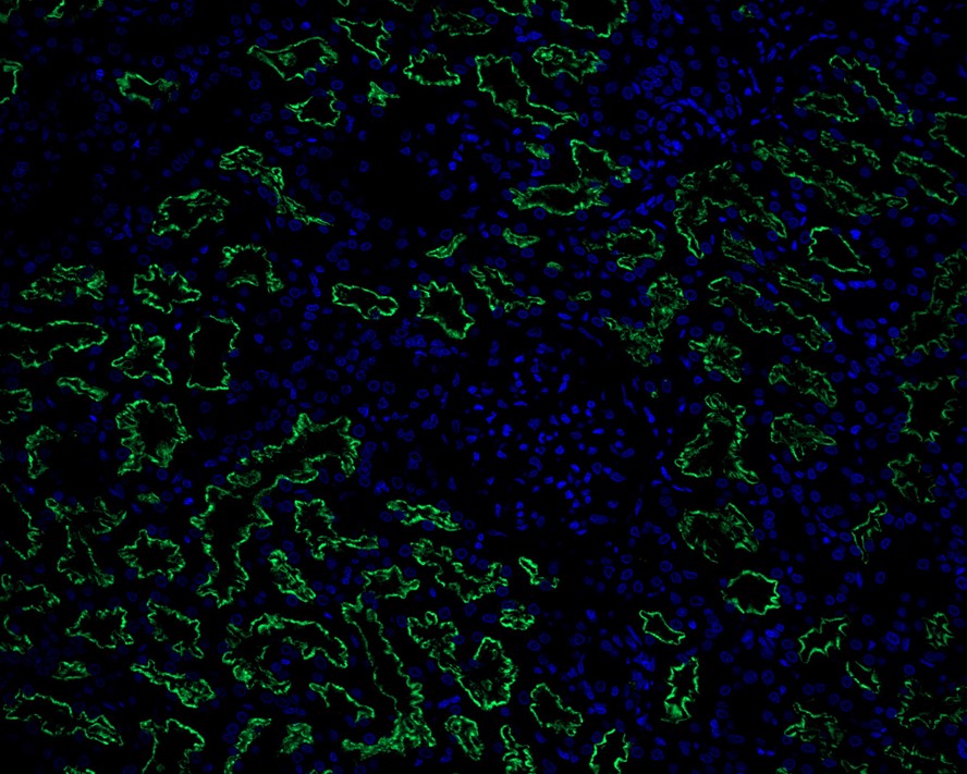
Immunofluorescence analysis of paraffin-embedded human liver tissue labeling Lrp2 / Megalin with Rabbit anti-Lrp2 / Megalin antibody at 1/50 dilution. The section was pre-treated using heat mediated antigen retrieval with Tris-EDTA buffer (pH 9.0) for 20 minutes. The tissues were blocked in 10% negative goat serum for 1 hour at room temperature, washed with PBS, and then probed with the primary antibody (green) at 1/50 dilution overnight at 4 ℃, washed with PBS. Goat Anti-Rabbit IgG H&L (488) was used as the secondary antibody at 1/1,000 dilution. Nuclei were counterstained with DAPI (blue).| Product Name | Lrp2 / Megalin Recombinant Rabbit Monoclonal Antibody |
|---|---|
| Antibody Type | Primary Antibodies |
| Immunogen | Synthetic peptide within human Lrp2 aa 2,500-3,200 / 4,655. |
| Clonality | Monoclonal |
|---|---|
| Isotype | IgG |
| Host Species | Rabbit |
| Tested Applications | IF-PIHCWB |
| IHC:1:1000 IF-P:1:50 WB:1:2000 |
|
| Species Reactivity | Human |
| Concentration | 1mg/ml |
| Purification | Protein A |
| Gene Symbol | LRP2 |
|---|---|
| Gene Synonyms | DBS GP330 LRP-2 |
| Gene Full Name | LDL receptor related protein 2 |
| Gene Summary | The protein encoded by this gene, low density lipoprotein-related protein 2 (LRP2) or megalin, is a multi-ligand endocytic receptor that is expressed in many different tissues but primarily in absorptive epithilial tissues such as the kidney. This glycoprotein has a large amino-terminal extracellular domain, a single transmembrane domain, and a short carboxy-terminal cytoplasmic tail. The extracellular ligand-binding-domains bind diverse macromolecules including albumin, apolipoproteins B and E, and lipoprotein lipase. The LRP2 protein is critical for the reuptake of numerous ligands, including lipoproteins, sterols, vitamin-binding proteins, and hormones. This protein also has a role in cell-signaling; extracellular ligands include parathyroid horomones and the morphogen sonic hedgehog while cytosolic ligands include MAP kinase scaffold proteins and JNK interacting proteins. Recycling of this membrane receptor is regulated by phosphorylation of its cytoplasmic domain. Mutations in this gene cause Donnai-Barrow syndrome (DBS) and facio-oculoacoustico-renal syndrome (FOAR).[provided by RefSeq, Aug 2009] |
| Alternative Names | Calcium sensor protein antibody DBS antibody Glycoprotein 330 antibody gp330 antibody Heymann nephritis antigen homolog antibody Low-density lipoprotein receptor-related protein 2 antibody LRP-2 antibody Lrp2 antibody LRP2_HUMAN antibody Megalin antibody |
| Molecular Weight(MW) | 522 kDa(Observed band size: 600 kDa) |
| Cellular Localization | Apical cell membrane, Endosome lumen, Membrane, coated pit, Cell projection, dendrite, axon. |
WB
Western blot analysis of Lrp2 / Megalin on human kidney tissue lysates with Rabbit anti-Lrp2 / Megalin antibody at 1/2,000 dilution. Lysates/proteins at 40 µg/Lane. Exposure time: 2 minutes; 4-20% SDS-PAGE gel. Proteins were transferred to a PVDF membrane and blocked with 5% NFDM/TBST for 1 hour at room temperature. The primary antibody at 1/2,000 dilution was used in 5% NFDM/TBST at 4℃ overnight. Goat Anti-Rabbit IgG - HRP Secondary Antibody at 1/50,000 dilution was used for 1 hour at room temperature.
IHC
Immunohistochemical analysis of paraffin-embedded human kidney tissue with Rabbit anti-Lrp2 / Megalin antibody at 1/1000 dilution. The section was pre-treated using heat mediated antigen retrieval with Tris-EDTA buffer (pH 9.0) for 20 minutes. The tissues were blocked in 1% BSA for 20 minutes at room temperature, washed with ddH2O and PBS, and then probed with the primary antibody at 1/200 dilution for 1 hour at room temperature. The detection was performed using an HRP conjugated compact polymer system. DAB was used as the chromogen. Tissues were counterstained with hematoxylin and mounted with DPX.
IHC
Immunohistochemical analysis of paraffin-embedded human skeletal muscle tissue (negative) with Rabbit anti-Lrp2 / Megalin antibody at 1/1000 dilution. The section was pre-treated using heat mediated antigen retrieval with Tris-EDTA buffer (pH 9.0) for 20 minutes. The tissues were blocked in 1% BSA for 20 minutes at room temperature, washed with ddH2O and PBS, and then probed with the primary antibody at 1/200 dilution for 1 hour at room temperature. The detection was performed using an HRP conjugated compact polymer system. DAB was used as the chromogen. Tissues were counterstained with hematoxylin and mounted with DPX.
IF-P
Immunofluorescence analysis of paraffin-embedded human liver tissue labeling Lrp2 / Megalin with Rabbit anti-Lrp2 / Megalin antibody at 1/50 dilution. The section was pre-treated using heat mediated antigen retrieval with Tris-EDTA buffer (pH 9.0) for 20 minutes. The tissues were blocked in 10% negative goat serum for 1 hour at room temperature, washed with PBS, and then probed with the primary antibody (green) at 1/50 dilution overnight at 4 ℃, washed with PBS. Goat Anti-Rabbit IgG H&L (488) was used as the secondary antibody at 1/1,000 dilution. Nuclei were counterstained with DAPI (blue).| Application Notes | IHC:1:1000 IF-P:1:50 WB:1:2000 |
|---|
| Form | Liquid |
|---|---|
| Storage Instructions | Shipped at 4℃. Store at +4℃ short term (1-2 weeks). It is recommended to aliquot into single-use upon delivery. Store at -20℃ long term. |
| Storage Buffer | PBS (pH7.4), 0.1% BSA, 40% Glycerol. Preservative: 0.05% Sodium Azide. |


