IF-F
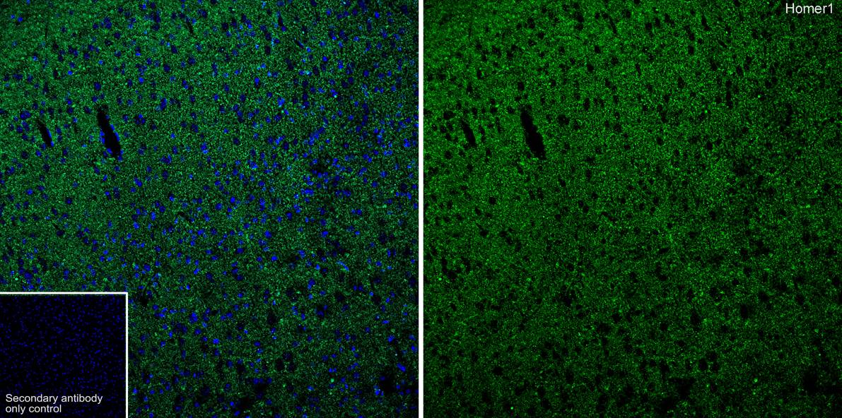
Application: IHC-Fr; Species: Mouse; Site: Cerebral cortex; Sample: Frozen section; Antibody concentration: 1:500; Antigen retrieval: Not required.IF-F
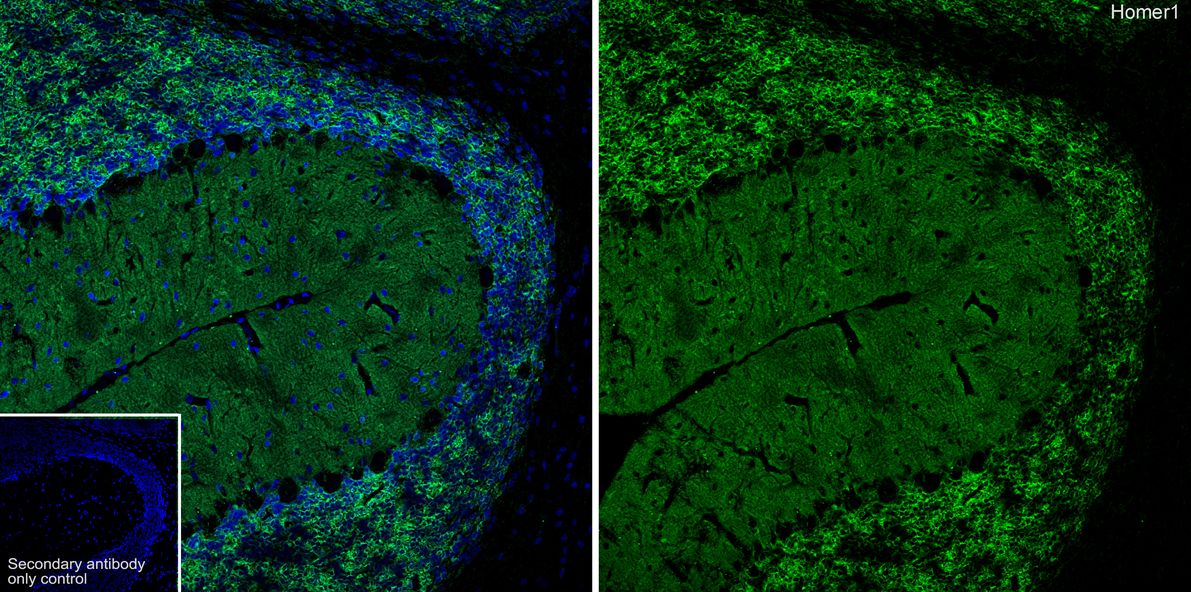
Application: IHC-Fr; Species: Rat; Site: Cerebellum; Sample: Frozen section; Antibody concentration: 1:500; Antigen retrieval: Not required.WB
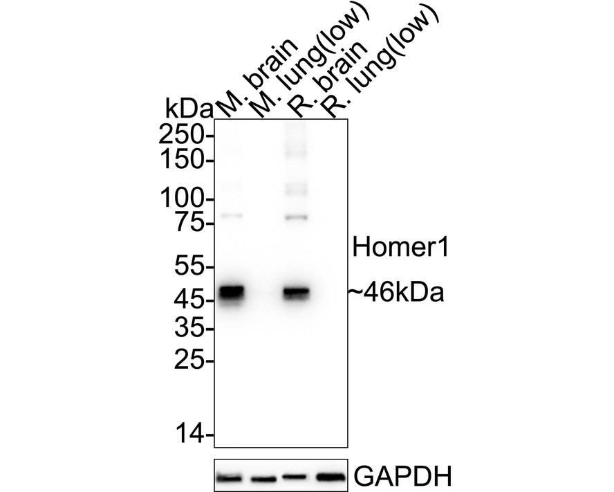
Western blot analysis of Homer1 on different lysates with Rabbit anti-Homer1 antibody at 1/20,000 dilution. Exposure time: 8 seconds; 4-20% SDS-PAGE gel. Proteins were transferred to a PVDF membrane and blocked with 5% NFDM/TBST for 1 hour at room temperature. The primary antibody at 1/20,000 dilution was used in primary antibody dilution at 4℃ overnight. Goat Anti-Rabbit IgG - HRP Secondary Antibody at 1/50,000 dilution was used for 1 hour at room temperature. Low expression: heart, kidney and lung.IHC
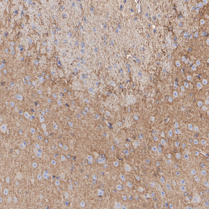
Immunohistochemical analysis of paraffin-embedded mouse brain tissue with Rabbit anti-Homer1 antibody at 1/400 dilution. The section was pre-treated using heat mediated antigen retrieval with Tris-EDTA buffer (pH 9.0) for 20 minutes. The tissues were blocked in 1% BSA for 20 minutes at room temperature, washed with ddH2O and PBS, and then probed with the primary antibody at 1/400 dilution for 1 hour at room temperature. The detection was performed using an HRP conjugated compact polymer system. DAB was used as the chromogen. Tissues were counterstained with hematoxylin and mounted with DPX.IHC
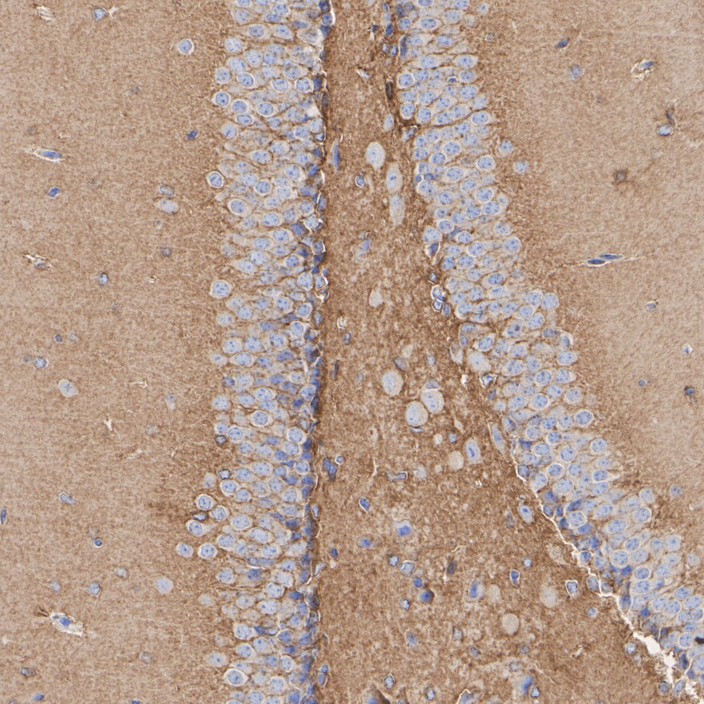
Immunohistochemical analysis of paraffin-embedded mouse hippocampus tissue with Rabbit anti-Homer1 antibody at 1/400 dilution. The section was pre-treated using heat mediated antigen retrieval with Tris-EDTA buffer (pH 9.0) for 20 minutes. The tissues were blocked in 1% BSA for 20 minutes at room temperature, washed with ddH2O and PBS, and then probed with the primary antibody at 1/400 dilution for 1 hour at room temperature. The detection was performed using an HRP conjugated compact polymer system. DAB was used as the chromogen. Tissues were counterstained with hematoxylin and mounted with DPX.IHC
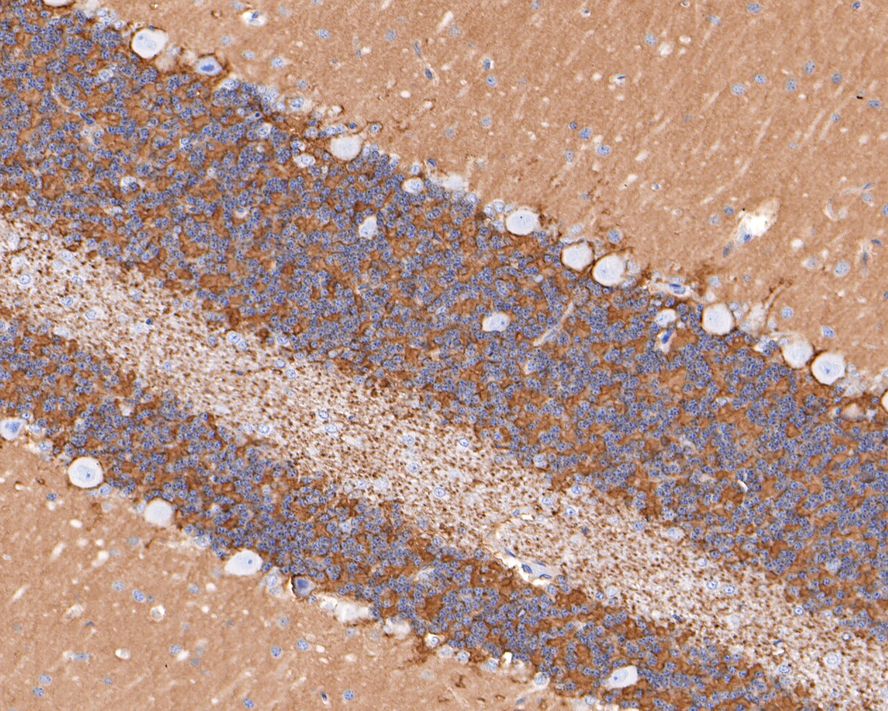
Immunohistochemical analysis of paraffin-embedded rat cerebellum tissue with Rabbit anti-Homer1 antibody at 1/500 dilution. The section was pre-treated using heat mediated antigen retrieval with Tris-EDTA buffer (pH 9.0) for 20 minutes. The tissues were blocked in 1% BSA for 20 minutes at room temperature, washed with ddH2O and PBS, and then probed with the primary antibody at 1/400 dilution for 1 hour at room temperature. The detection was performed using an HRP conjugated compact polymer system. DAB was used as the chromogen. Tissues were counterstained with hematoxylin and mounted with DPX.| Product Name | Homer1 Recombinant Rabbit Monoclonal Antibody |
|---|---|
| Antibody Type | Primary Antibodies |
| Immunogen | Recombinant protein within human Homer1 aa 101-250/354. |
| Clonality | Monoclonal |
|---|---|
| Isotype | IgG |
| Host Species | Rabbit |
| Tested Applications | IF-FIHCWB |
| WB:1:20000 IHC:1:500 IF-P:1:500 |
|
| Species Reactivity | Cynomolgus MonkeyMousePigRat |
| Concentration | 1mg/ml |
| Purification | Protein A |
| Gene Symbol | Homer1 |
|---|---|
| Gene Synonyms | SYN47 Ves-1 vesl-1 homer-1 PSD-Zip45 |
| Gene Full Name | homer scaffolding protein 1 |
| Gene Summary | Predicted to enable several functions, including scaffold protein binding activity; transmembrane transporter binding activity; and type 5 metabotropic glutamate receptor binding activity. Predicted to be a structural constituent of postsynapse. Involved in regulation of monoatomic ion transport and regulation of postsynaptic neurotransmitter receptor activity. Acts upstream of or within several processes, including behavioral response to cocaine; skeletal muscle contraction; and skeletal muscle fiber development. Located in several cellular components, including apical part of cell; myofibril; and postsynapse. Is active in glutamatergic synapse; postsynaptic cytosol; and postsynaptic density. Is expressed in several structures, including alimentary system; central nervous system; genitourinary system; respiratory system; and sensory organ. Orthologous to human HOMER1 (homer scaffold protein 1). [provided by Alliance of Genome Resources, Apr 2025] |
| Alternative Names | HOME1_HUMAN antibody Homer antibody HOMER 1A antibody HOMER 1B antibody HOMER 1C antibody Homer homolog 1 (Drosophila) antibody homer homolog 1 antibody Homer protein homolog 1 antibody homer scaffolding protein 1 antibody Homer Drosophila homolog of 1 antibody HOME1_HUMAN antibody Homer antibody HOMER 1A antibody HOMER 1B antibody HOMER 1C antibody Homer homolog 1 (Drosophila) antibody homer homolog 1 antibody Homer protein homolog 1 antibody homer scaffolding protein 1 antibody Homer Drosophila homolog of 1 antibody Homer neuronal immediate early gene 1 antibody Homer-1 antibody Homer1 antibody HOMER1A antibody HOMER1B antibody HOMER1C antibody SYN47 antibody Ves 1 antibody |
| Molecular Weight(MW) | 40 kDa(Observed band size: 46 kDa) |
| Cellular Localization | Cytoplasm, postsynaptic density, synapse, dendritic spine. |
IF-F
Application: IHC-Fr; Species: Mouse; Site: Cerebral cortex; Sample: Frozen section; Antibody concentration: 1:500; Antigen retrieval: Not required.
IF-F
Application: IHC-Fr; Species: Rat; Site: Cerebellum; Sample: Frozen section; Antibody concentration: 1:500; Antigen retrieval: Not required.
WB
Western blot analysis of Homer1 on different lysates with Rabbit anti-Homer1 antibody at 1/20,000 dilution. Exposure time: 8 seconds; 4-20% SDS-PAGE gel. Proteins were transferred to a PVDF membrane and blocked with 5% NFDM/TBST for 1 hour at room temperature. The primary antibody at 1/20,000 dilution was used in primary antibody dilution at 4℃ overnight. Goat Anti-Rabbit IgG - HRP Secondary Antibody at 1/50,000 dilution was used for 1 hour at room temperature. Low expression: heart, kidney and lung.
IHC
Immunohistochemical analysis of paraffin-embedded mouse brain tissue with Rabbit anti-Homer1 antibody at 1/400 dilution. The section was pre-treated using heat mediated antigen retrieval with Tris-EDTA buffer (pH 9.0) for 20 minutes. The tissues were blocked in 1% BSA for 20 minutes at room temperature, washed with ddH2O and PBS, and then probed with the primary antibody at 1/400 dilution for 1 hour at room temperature. The detection was performed using an HRP conjugated compact polymer system. DAB was used as the chromogen. Tissues were counterstained with hematoxylin and mounted with DPX.
IHC
Immunohistochemical analysis of paraffin-embedded mouse hippocampus tissue with Rabbit anti-Homer1 antibody at 1/400 dilution. The section was pre-treated using heat mediated antigen retrieval with Tris-EDTA buffer (pH 9.0) for 20 minutes. The tissues were blocked in 1% BSA for 20 minutes at room temperature, washed with ddH2O and PBS, and then probed with the primary antibody at 1/400 dilution for 1 hour at room temperature. The detection was performed using an HRP conjugated compact polymer system. DAB was used as the chromogen. Tissues were counterstained with hematoxylin and mounted with DPX.
IHC
Immunohistochemical analysis of paraffin-embedded rat cerebellum tissue with Rabbit anti-Homer1 antibody at 1/500 dilution. The section was pre-treated using heat mediated antigen retrieval with Tris-EDTA buffer (pH 9.0) for 20 minutes. The tissues were blocked in 1% BSA for 20 minutes at room temperature, washed with ddH2O and PBS, and then probed with the primary antibody at 1/400 dilution for 1 hour at room temperature. The detection was performed using an HRP conjugated compact polymer system. DAB was used as the chromogen. Tissues were counterstained with hematoxylin and mounted with DPX.| Application Notes | WB:1:20000 IHC:1:500 IF-P:1:500 |
|---|
| Form | Liquid |
|---|---|
| Storage Instructions | Shipped at 4℃. Store at +4℃ short term (1-2 weeks). It is recommended to aliquot into single-use upon delivery. Store at -20℃ long term. |
| Storage Buffer | TBS (pH7.4), 0.05% BSA, 40% Glycerol. Preservative: 0.05% Sodium Azide. |


