WB
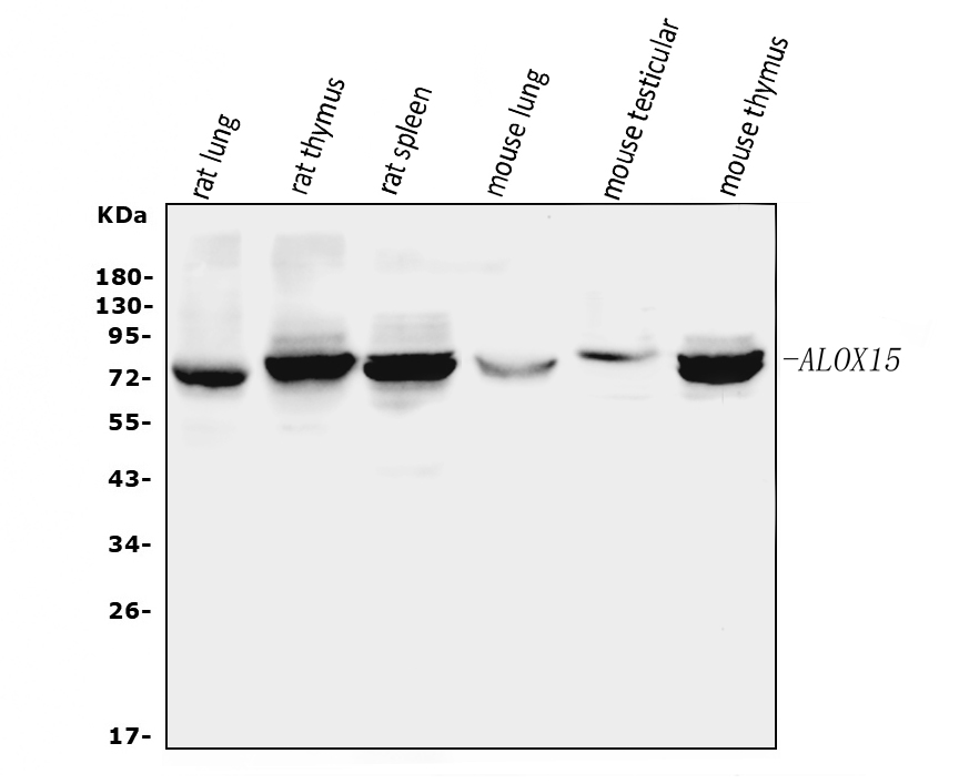
Western blot analysis of ALOX15 using anti-ALOX15 antibody. The sample well of each lane was loaded with 30 ug of sample under reducing conditions. After electrophoresis, proteins were transferred to a membrane. Then the membrane was incubated with rabbit anti-ALOX15 antigen affinity purified polyclonal antibody at a dilution of 1:1000 and probed with a goat anti-rabbit IgG-HRP secondary antibody. The signal is developed using ECL Plus Western Blotting Substrate.IHC
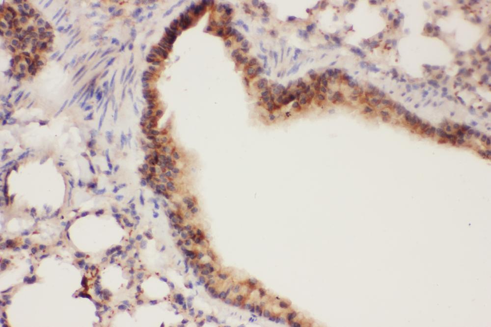
IHC analysis of ALOX15 using anti-ALOX15 antibody. ALOX15 was detected in a paraffin-embedded section of Mouse Lung tissue. Biotinylated goat anti-rabbit IgG was used as secondary antibody. The tissue section was incubated with rabbit anti-ALOX15 Antibody at a dilution of 1:200 and developed using Strepavidin-Biotin-Complex (SABC) with DAB as the chromogen.IHC
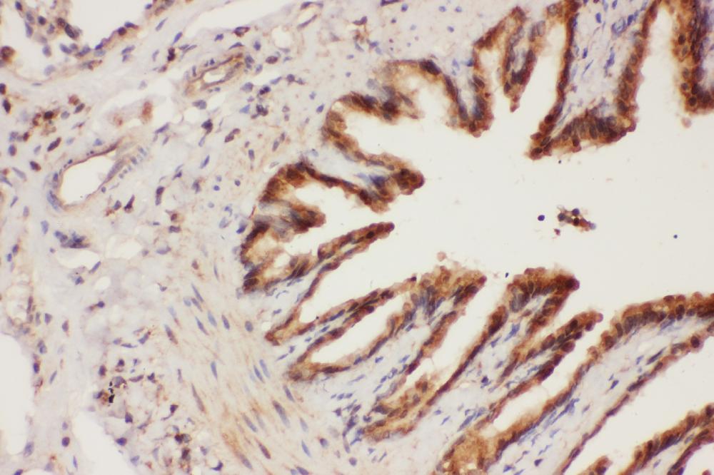
IHC analysis of ALOX15 using anti-ALOX15 antibody. ALOX15 was detected in a paraffin-embedded section of rat lung tissue. Biotinylated goat anti-rabbit IgG was used as secondary antibody. The tissue section was incubated with rabbit anti-ALOX15 Antibody at a dilution of 1:200 and developed using Strepavidin-Biotin-Complex (SABC) with DAB as the chromogen.IHC
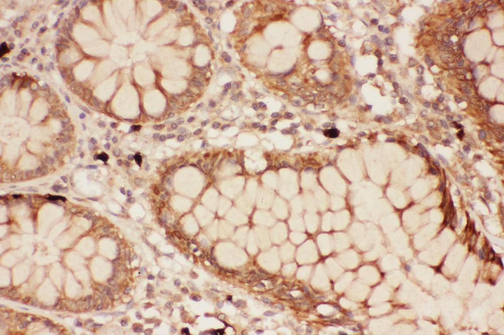
IHC analysis of ALOX15 using anti-ALOX15 antibody. ALOX15 was detected in a paraffin-embedded section of Human Intestine Cancer tissue. Biotinylated goat anti-rabbit IgG was used as secondary antibody. The tissue section was incubated with rabbit anti-ALOX15 Antibody at a dilution of 1:200 and developed using Strepavidin-Biotin-Complex (SABC) with DAB as the chromogen.IHC
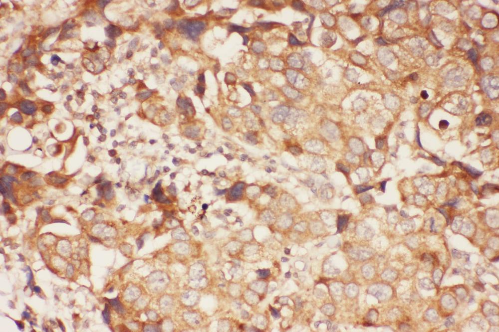
IHC analysis of ALOX15 using anti-ALOX15 antibody. ALOX15 was detected in a paraffin-embedded section of human mammary cancer tissue. Biotinylated goat anti-rabbit IgG was used as secondary antibody. The tissue section was incubated with rabbit anti-ALOX15 Antibody at a dilution of 1:200 and developed using Strepavidin-Biotin-Complex (SABC) with DAB as the chromogen.| Product Name | Rabbit polyclonal antibody to ALOX15 |
|---|---|
| Antibody Type | Primary Antibodies |
| Immunogen | E.coli-derived human ALOX15 recombinant protein (Position: G2-P337). Human ALOX15 shares 72% and 73% amino acid (aa) sequences identity with mouse and rat ALOX15, respectively. |
| Clonality | Polyclonal |
|---|---|
| Isotype | IgG |
| Host Species | Rabbit |
| Tested Applications | IHCWB |
| WB:1:500-1:2000 IHC:1:50-1:400 |
|
| Species Reactivity | HumanMouseRat |
| Concentration | 0.5mg/ml |
| Purification | Affinity purified |
| Gene Symbol | ALOX15 |
|---|---|
| Gene Synonyms | LOG15 12-LOX 15-LOX 15-LOX-1 |
| Gene Full Name | arachidonate 15-lipoxygenase |
| Gene Summary | This gene encodes a member of the lipoxygenase family of proteins. The encoded enzyme acts on various polyunsaturated fatty acid substrates to generate various bioactive lipid mediators such as eicosanoids, hepoxilins, lipoxins, and other molecules. The encoded enzyme and its reaction products have been shown to regulate inflammation and immunity. Multiple pseudogenes of this gene have been identified in the human genome. [provided by RefSeq, Aug 2017] |
| Alternative Names | 12/15-lipoxygenase 12-LOX Arachidonate 15-lipoxygenase 15-LOX 15-LOX-1 Arachidonate omega-6 lipoxygenase Hepoxilin A3 synthase Alox15 Linoleate 13S-lipoxygenase ALOX15 LOG15 12-LOX |
| Molecular Weight(MW) | 75 kDa |
| Cellular Localization | Cell membrane,Cytoplasm,Lipid droplet,Membrane |
WB
Western blot analysis of ALOX15 using anti-ALOX15 antibody. The sample well of each lane was loaded with 30 ug of sample under reducing conditions. After electrophoresis, proteins were transferred to a membrane. Then the membrane was incubated with rabbit anti-ALOX15 antigen affinity purified polyclonal antibody at a dilution of 1:1000 and probed with a goat anti-rabbit IgG-HRP secondary antibody. The signal is developed using ECL Plus Western Blotting Substrate.
IHC
IHC analysis of ALOX15 using anti-ALOX15 antibody. ALOX15 was detected in a paraffin-embedded section of Mouse Lung tissue. Biotinylated goat anti-rabbit IgG was used as secondary antibody. The tissue section was incubated with rabbit anti-ALOX15 Antibody at a dilution of 1:200 and developed using Strepavidin-Biotin-Complex (SABC) with DAB as the chromogen.
IHC
IHC analysis of ALOX15 using anti-ALOX15 antibody. ALOX15 was detected in a paraffin-embedded section of rat lung tissue. Biotinylated goat anti-rabbit IgG was used as secondary antibody. The tissue section was incubated with rabbit anti-ALOX15 Antibody at a dilution of 1:200 and developed using Strepavidin-Biotin-Complex (SABC) with DAB as the chromogen.
IHC
IHC analysis of ALOX15 using anti-ALOX15 antibody. ALOX15 was detected in a paraffin-embedded section of Human Intestine Cancer tissue. Biotinylated goat anti-rabbit IgG was used as secondary antibody. The tissue section was incubated with rabbit anti-ALOX15 Antibody at a dilution of 1:200 and developed using Strepavidin-Biotin-Complex (SABC) with DAB as the chromogen.
IHC
IHC analysis of ALOX15 using anti-ALOX15 antibody. ALOX15 was detected in a paraffin-embedded section of human mammary cancer tissue. Biotinylated goat anti-rabbit IgG was used as secondary antibody. The tissue section was incubated with rabbit anti-ALOX15 Antibody at a dilution of 1:200 and developed using Strepavidin-Biotin-Complex (SABC) with DAB as the chromogen.| Application Notes | WB:1:500-1:2000 IHC:1:50-1:400 |
|---|
| Form | Liquid |
|---|---|
| Storage Instructions | 12 months from date of receipt, -20℃ as supplied. 6 months 2 to 8℃ after reconstitution. Avoid repeated freezing and thawing. |
| Storage Buffer | 500ug/ml antibody with PBS, 0.02% NaN3, 1 mg/ml BSA and 50% glycerol. |


