WB
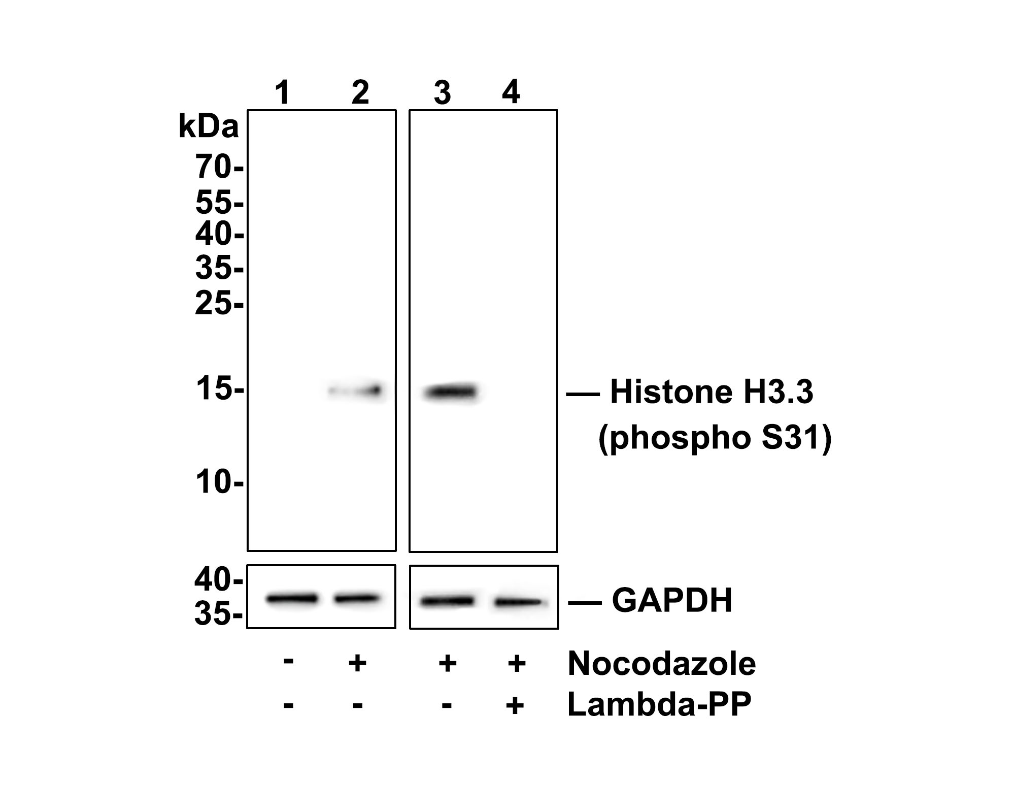
Western blot analysis of Histone H3.3 (phospho S31) on Hela cell lysates. Lane 1: Hela cells, whole cell lysate, 10ug/lane Lane 2/3: Hela cells treated with 100ng/ml Nocodazole for 18h, whole cell lysates, 10ug/lane Lane 4: Hela cells treated with 100ng/ml Nocodazole for 18h, then treated with 2.8ug/ul lambda-PP for 30 minutes, whole cell lysates, 10ug/lane All lanes : Anti-Histone H3.3 (phospho S31) antibody at 1:500 dilution. Anti-GAPDH antibody at 1:10,000 dilution. Goat Anti-Rabbit IgG H&L (HRP) at 1/200,000 dilution. Blocking and diluting buffer: 5% BSA. Exposure time: 2 minutes 34 seconds Proteins were transferred to a PVDF membrane and blocked with 5% NFDM/TBST for 1 hour at room temperature. The primary antibody at 1/500 dilution was used in 5% NFDM/TBST at room temperature for 2 hours. Goat Anti-Rabbit IgG - HRP Secondary Antibody at 1:200,000 dilution was used for 1 hour at room temperature.WB
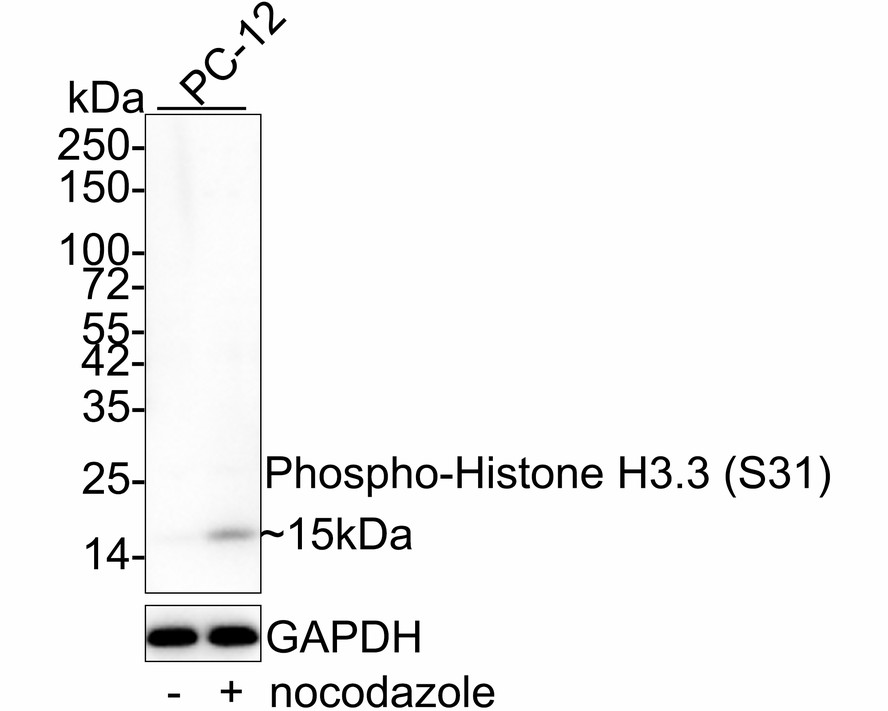
Western blot analysis of Phospho-Histone H3.3 (S31) on different lysates with Rabbit anti-Phospho-Histone H3.3 (S31) antibody at 1/1,000 dilution. Exposure time: 1 minute; 4-20% SDS-PAGE gel. Proteins were transferred to a PVDF membrane and blocked with 5% NFDM/TBST for 1 hour at room temperature. The primary antibody at 1/1,000 dilution was used in 5% NFDM/TBST at 4℃ overnight. Goat Anti-Rabbit IgG - HRP Secondary Antibody at 1/50,000 dilution was used for 1 hour at room temperature.ICC/IF
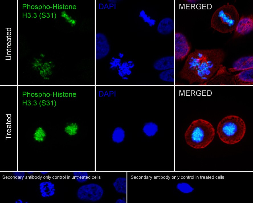
Immunocytochemistry analysis of HeLa cells treated with or without 100ng/mL Nocodazole for 18 hours labeling Phospho-Histone H3.3 (S31) with Rabbit anti-Phospho-Histone H3.3 (S31) antibody at 1/25,000 dilution. Cells were fixed in 4% paraformaldehyde for 20 minutes at room temperature, permeabilized with 0.1% Triton X-100 in PBS for 5 minutes at room temperature, then blocked with 1% BSA in 10% negative goat serum for 1 hour at room temperature. Cells were then incubated with Rabbit anti-Phospho-Histone H3.3 (S31) antibody at 1/25,000 dilution in 1% BSA in PBST overnight at 4 ℃. Goat Anti-Rabbit IgG H&L (488) was used as the secondary antibody at 1/1,000 dilution. PBS instead of the primary antibody was used as the secondary antibody only control. Nuclear DNA was labelled in blue with DAPI. Beta tubulin (red) was stained at 1/100 dilution overnight at +4℃. Goat Anti-Mouse IgG H&L (594) was used as the secondary antibody at 1/1,000 dilution.| Product Name | Phospho-Histone H3.3 (S31) Recombinant Rabbit Monoclonal Antibody |
|---|---|
| Antibody Type | Primary Antibodies |
| Immunogen | Synthetic phospho-peptide corresponding to residues surrounding Ser31 of Human Histone H3.3. |
| Modification | p-S31 |
| Clonality | Monoclonal |
|---|---|
| Isotype | IgG |
| Host Species | Rabbit |
| Tested Applications | ICC/IFWB |
| WB:1:500-1:1000 ICC/IF:1:25000 |
|
| Species Reactivity | HumanRat |
| Concentration | 1mg/ml |
| Purification | Protein A |
| Gene Symbol | H3-3A |
|---|---|
| Gene Synonyms | H3F3 H3.3A H3F3A BRYLIB1 |
| Gene Full Name | H3.3 histone A |
| Gene Summary | Histones are basic nuclear proteins that are responsible for the nucleosome structure of the chromosomal fiber in eukaryotes. Two molecules of each of the four core histones (H2A, H2B, H3, and H4) form an octamer, around which approximately 146 bp of DNA is wrapped in repeating units, called nucleosomes. The linker histone, H1, interacts with linker DNA between nucleosomes and functions in the compaction of chromatin into higher order structures. This gene contains introns and its mRNA is polyadenylated, unlike most histone genes. The protein encoded is a replication-independent member of the histone H3 family. [provided by RefSeq, Jul 2008] |
| Molecular Weight(MW) | 15 kDa |
| Cellular Localization | Nucleus, Chromosome. |
| SwissProt ID | P84243 |
|---|
WB
Western blot analysis of Histone H3.3 (phospho S31) on Hela cell lysates. Lane 1: Hela cells, whole cell lysate, 10ug/lane Lane 2/3: Hela cells treated with 100ng/ml Nocodazole for 18h, whole cell lysates, 10ug/lane Lane 4: Hela cells treated with 100ng/ml Nocodazole for 18h, then treated with 2.8ug/ul lambda-PP for 30 minutes, whole cell lysates, 10ug/lane All lanes : Anti-Histone H3.3 (phospho S31) antibody at 1:500 dilution. Anti-GAPDH antibody at 1:10,000 dilution. Goat Anti-Rabbit IgG H&L (HRP) at 1/200,000 dilution. Blocking and diluting buffer: 5% BSA. Exposure time: 2 minutes 34 seconds Proteins were transferred to a PVDF membrane and blocked with 5% NFDM/TBST for 1 hour at room temperature. The primary antibody at 1/500 dilution was used in 5% NFDM/TBST at room temperature for 2 hours. Goat Anti-Rabbit IgG - HRP Secondary Antibody at 1:200,000 dilution was used for 1 hour at room temperature.
WB
Western blot analysis of Phospho-Histone H3.3 (S31) on different lysates with Rabbit anti-Phospho-Histone H3.3 (S31) antibody at 1/1,000 dilution. Exposure time: 1 minute; 4-20% SDS-PAGE gel. Proteins were transferred to a PVDF membrane and blocked with 5% NFDM/TBST for 1 hour at room temperature. The primary antibody at 1/1,000 dilution was used in 5% NFDM/TBST at 4℃ overnight. Goat Anti-Rabbit IgG - HRP Secondary Antibody at 1/50,000 dilution was used for 1 hour at room temperature.
ICC/IF
Immunocytochemistry analysis of HeLa cells treated with or without 100ng/mL Nocodazole for 18 hours labeling Phospho-Histone H3.3 (S31) with Rabbit anti-Phospho-Histone H3.3 (S31) antibody at 1/25,000 dilution. Cells were fixed in 4% paraformaldehyde for 20 minutes at room temperature, permeabilized with 0.1% Triton X-100 in PBS for 5 minutes at room temperature, then blocked with 1% BSA in 10% negative goat serum for 1 hour at room temperature. Cells were then incubated with Rabbit anti-Phospho-Histone H3.3 (S31) antibody at 1/25,000 dilution in 1% BSA in PBST overnight at 4 ℃. Goat Anti-Rabbit IgG H&L (488) was used as the secondary antibody at 1/1,000 dilution. PBS instead of the primary antibody was used as the secondary antibody only control. Nuclear DNA was labelled in blue with DAPI. Beta tubulin (red) was stained at 1/100 dilution overnight at +4℃. Goat Anti-Mouse IgG H&L (594) was used as the secondary antibody at 1/1,000 dilution.| Application Notes | WB:1:500-1:1000 ICC/IF:1:25000 |
|---|
| Form | Liquid |
|---|---|
| Storage Instructions | Shipped at 4°C. Store at +4°C short term (1-2 weeks). Store at -20°C long term. Avoid freeze / thaw cycle. |
| Storage Buffer | TBS (pH7.4), 0.05% BSA, 40% Glycerol. Preservative: 0.05% Sodium Azide. |


