WB
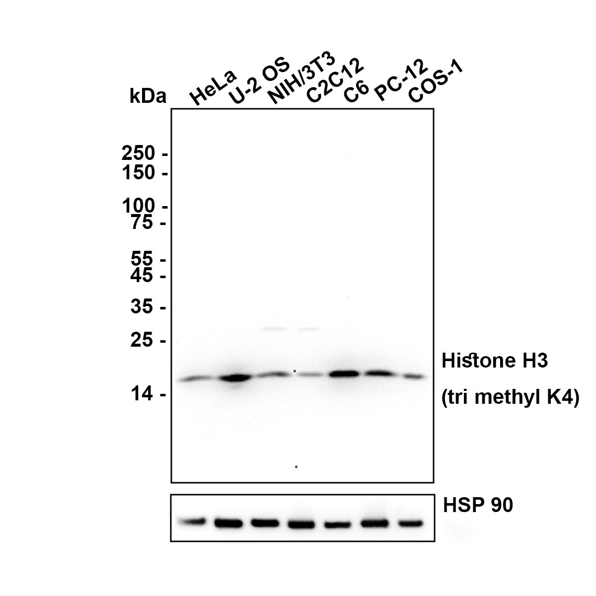
Western blot analysis of Histone H3 (tri methyl K4) on different lysates with Rabbit anti-Histone H3 (tri methyl K4) antibody at 1/2,000 dilution. Lysates/proteins at 20 µg/Lane. Exposure time: 1 minute 30 seconds; Blocking: 5% NFDM/TBST, 1 hour at room temperature Primary antibody: 1/2,000 in primary antibody dilution buffer, overnight at 4 °C. Secondary antibody: Goat anti-Rabbit IgG-HRP, 1/50,000 in 5% NFDM/TBST, 1 hour at room temperature.ICC/IF
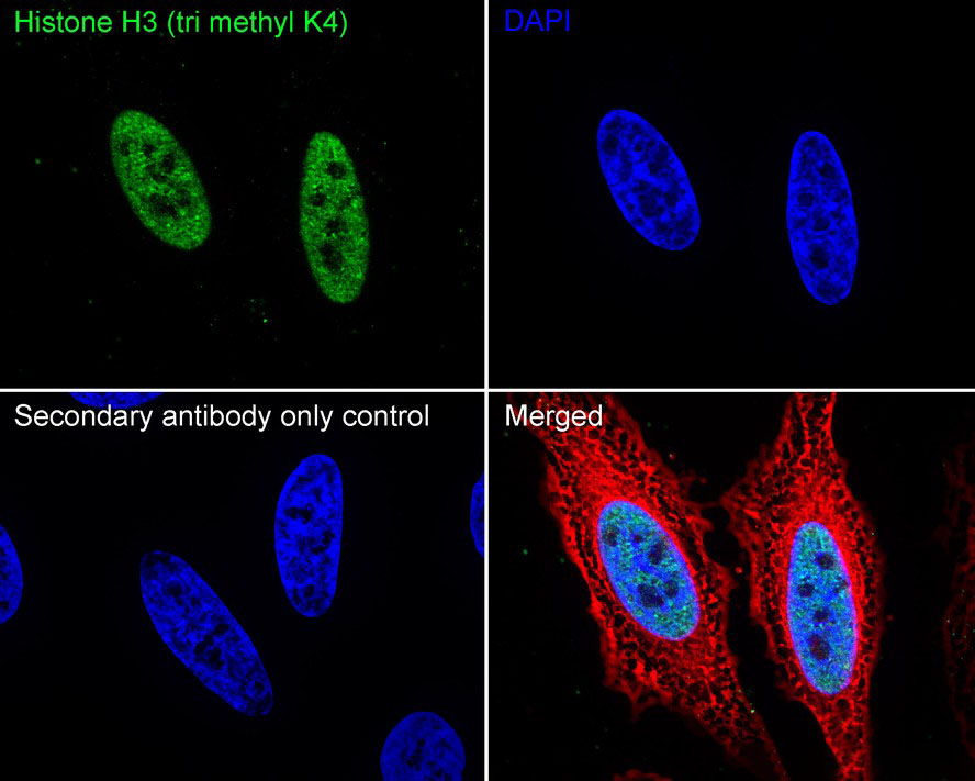
Application: Immunocytochemistry (IF-cell) Species: Human Sample: HeLa (Human cervical adenocarcinoma cell) Fixation: 4% Paraformaldehyde, 15 minutes at room temperature. Permeabilization: 0.1% Triton X-100, 15 minutes at room temperature. Blocking: 1% BSA + 10% normal goat serum, 1 hour at room temperature. Antibody dilution buffer: 1% BSA in PBST. Primary antibody: 1/500, overnight at 4°C. Secondary antibody: Goat Anti-Rabbit IgG (488), 45 minutes at room temperature. Counterstain: Beta tubulin (Red), 1/100, overnight at 4℃. The nuclear counterstain was DAPI (Blue).ICC/IF
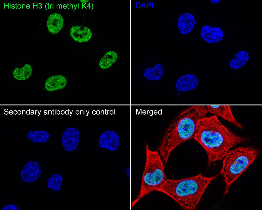
Application: Immunocytochemistry (IF-cell) Species: Mouse Sample: NIH/3T3 (Mouse fibroblast) Fixation: 4% Paraformaldehyde, 15 minutes at room temperature. Permeabilization: 0.1% Triton X-100, 15 minutes at room temperature. Blocking: 1% BSA + 10% normal goat serum, 1 hour at room temperature. Antibody dilution buffer: 1% BSA in PBST. Primary antibody: 1/500, overnight at 4°C. Secondary antibody: Goat Anti-Rabbit IgG (488), 45 minutes at room temperature. Counterstain: Beta tubulin (Red), 1/100, overnight at 4℃. The nuclear counterstain was DAPI (Blue).ICC/IF
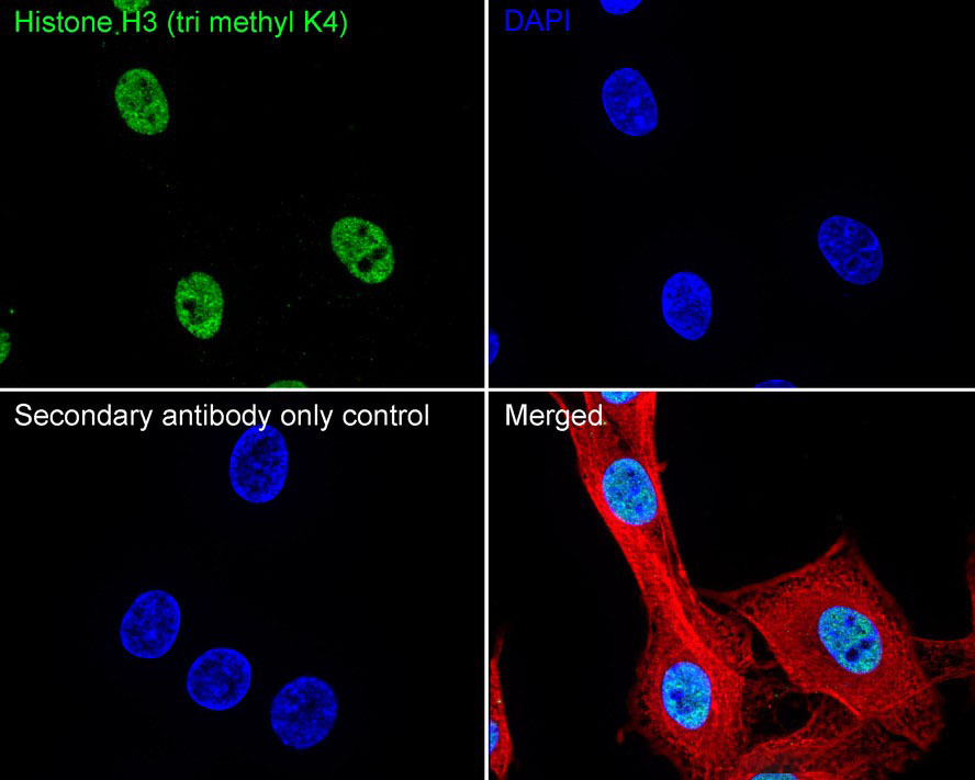
Application: Immunocytochemistry (IF-cell) Species: Rat Sample: C6 (Rat glioma cell) Fixation: 4% Paraformaldehyde, 15 minutes at room temperature. Permeabilization: 0.1% Triton X-100, 15 minutes at room temperature. Blocking: 1% BSA + 10% normal goat serum, 1 hour at room temperature. Antibody dilution buffer: 1% BSA in PBST. Primary antibody: 1/500, overnight at 4°C. Secondary antibody: Goat Anti-Rabbit IgG (488), 45 minutes at room temperature. Counterstain: Beta tubulin (Red), 1/100, overnight at 4℃. The nuclear counterstain was DAPI (Blue).IHC
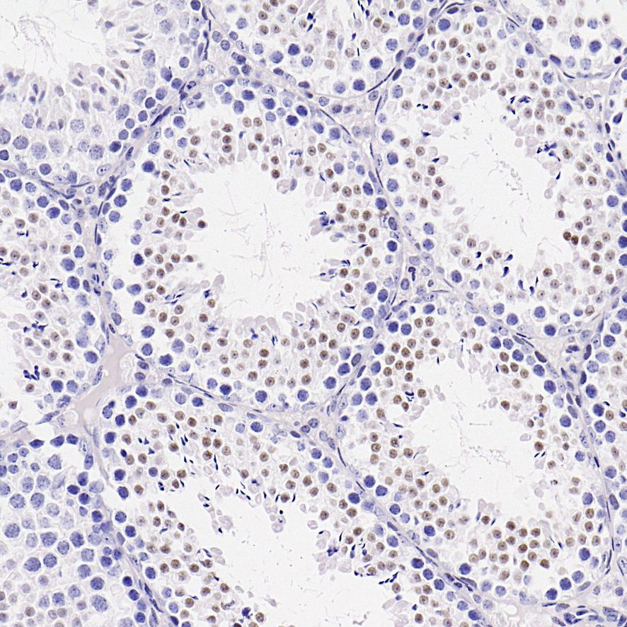
Application: Immunohistochemistry (IHC-P) Species: Mouse Tissue: Testis Sample: Paraffin-embedded section Antigen retrieval: Heat-mediated, Tris-EDTA buffer (pH 9.0), 20 minutes at 95°C. Wash buffer: 1× TBST Endogenous peroxidase blocking: 3% H₂O₂, 10 minutes at room temperature. Blocking: 1% BSA + 10% normal goat serum, 10 minutes at room temperature. Primary antibody: 1/100, 1 hour at room temperature. Secondary antibody: 20 minutes at room temperature.IHC
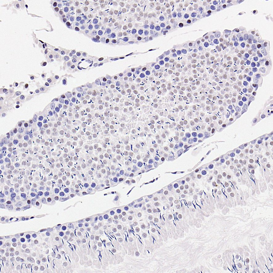
Application: Immunohistochemistry (IHC-P) Species: Rat Tissue: Testis Sample: Paraffin-embedded section Antigen retrieval: Heat-mediated, Tris-EDTA buffer (pH 9.0), 20 minutes at 95°C. Wash buffer: 1× TBST Endogenous peroxidase blocking: 3% H₂O₂, 10 minutes at room temperature. Blocking: 1% BSA + 10% normal goat serum, 10 minutes at room temperature. Primary antibody: 1/100, 1 hour at room temperature. Secondary antibody: 20 minutes at room temperature.FC
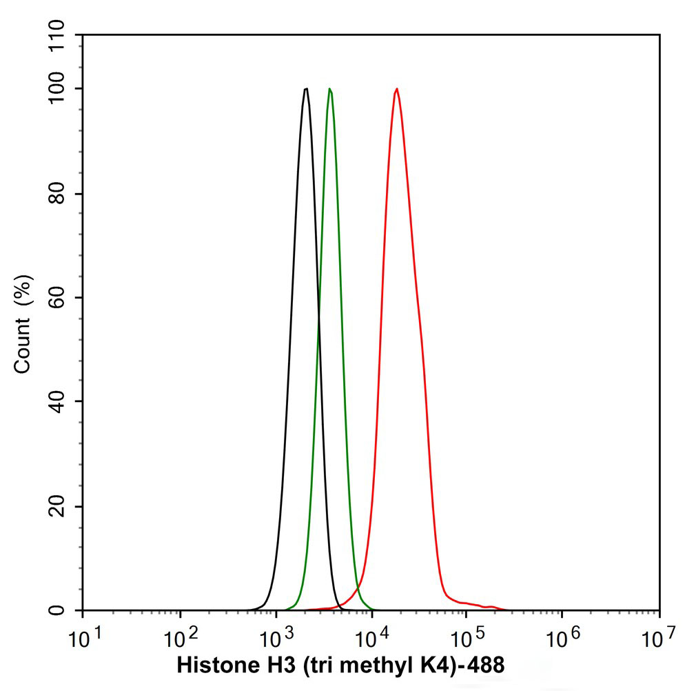
Application: Flow Cytometry (Intra) Species: Human Sample: HeLa (Human cervix adenocarcinoma epithelial cell) Fixation: 4% Paraformaldehyde, 15 minutes at room temperature. Permeabilization: 0.1% Tween-20, 15 minutes at room temperature. Blocking: 1% BSA + 10% normal goat serum, 15 minutes at room temperature. Antibody dilution buffer: 1x PBS. Primary antibody: (1/1,000, Red) compared with Rabbit IgG Isotype Control (Green), 15 minutes at room temperature. Secondary antibody: Goat Anti-Rabbit IgG (488), 15 minutes at room temperature.CHIP
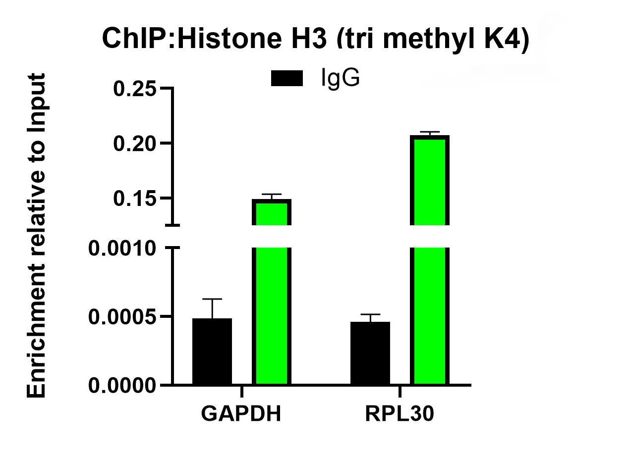
Chromatin immunoprecipitations were performed with cross-linked chromatin from HeLa cells with Histone H3 (tri methyl K4) / Competitor's antibody / Normal Rabbit IgG according to the ChIP protocol. The enriched DNA was quantified by real-time PCR using indicated primers. The amount of immunoprecipitated DNA in each sample is represented as signal relative to the total amount of input chromatin, which is equivalent to one.| Product Name | Histone H3 (tri methyl K4) Recombinant Rabbit Monoclonal Antibody |
|---|---|
| Antibody Type | Primary Antibodies |
| Clonality | Monoclonal |
|---|---|
| Isotype | IgG |
| Host Species | Rabbit |
| Tested Applications | ChIPFCICC/IFIHCWB |
| WB:1:2000 ICC/IF:1:500 IHC:1:50-1:100 FC(Intra):1:1000 ChIP:Use 0.5~2 μg for 25 μg of chromatin. |
|
| Species Reactivity | HumanMonkeyMouseRat |
| Concentration | 1mg/ml |
| Purification | Protein A |
| Gene Symbol | H3C1 |
|---|---|
| Gene Synonyms | H3/A H3FA HIST1H3A |
| Gene Full Name | H3 clustered histone 1 |
| Gene Summary | Histones are basic nuclear proteins that are responsible for the nucleosome structure of the chromosomal fiber in eukaryotes. This structure consists of approximately 146 bp of DNA wrapped around a nucleosome, an octamer composed of pairs of each of the four core histones (H2A, H2B, H3, and H4). The chromatin fiber is further compacted through the interaction of a linker histone, H1, with the DNA between the nucleosomes to form higher order chromatin structures. This gene is intronless and encodes a replication-dependent histone that is a member of the histone H3 family. Transcripts from this gene lack polyA tails; instead, they contain a palindromic termination element. This gene is found in the large histone gene cluster on chromosome 6p22-p21.3. [provided by RefSeq, Aug 2015] |
| Molecular Weight(MW) | 15 kDa |
| Cellular Localization | Nucleus. |
| SwissProt ID | P68431 |
|---|
WB
Western blot analysis of Histone H3 (tri methyl K4) on different lysates with Rabbit anti-Histone H3 (tri methyl K4) antibody at 1/2,000 dilution. Lysates/proteins at 20 µg/Lane. Exposure time: 1 minute 30 seconds; Blocking: 5% NFDM/TBST, 1 hour at room temperature Primary antibody: 1/2,000 in primary antibody dilution buffer, overnight at 4 °C. Secondary antibody: Goat anti-Rabbit IgG-HRP, 1/50,000 in 5% NFDM/TBST, 1 hour at room temperature.
ICC/IF
Application: Immunocytochemistry (IF-cell) Species: Human Sample: HeLa (Human cervical adenocarcinoma cell) Fixation: 4% Paraformaldehyde, 15 minutes at room temperature. Permeabilization: 0.1% Triton X-100, 15 minutes at room temperature. Blocking: 1% BSA + 10% normal goat serum, 1 hour at room temperature. Antibody dilution buffer: 1% BSA in PBST. Primary antibody: 1/500, overnight at 4°C. Secondary antibody: Goat Anti-Rabbit IgG (488), 45 minutes at room temperature. Counterstain: Beta tubulin (Red), 1/100, overnight at 4℃. The nuclear counterstain was DAPI (Blue).
ICC/IF
Application: Immunocytochemistry (IF-cell) Species: Mouse Sample: NIH/3T3 (Mouse fibroblast) Fixation: 4% Paraformaldehyde, 15 minutes at room temperature. Permeabilization: 0.1% Triton X-100, 15 minutes at room temperature. Blocking: 1% BSA + 10% normal goat serum, 1 hour at room temperature. Antibody dilution buffer: 1% BSA in PBST. Primary antibody: 1/500, overnight at 4°C. Secondary antibody: Goat Anti-Rabbit IgG (488), 45 minutes at room temperature. Counterstain: Beta tubulin (Red), 1/100, overnight at 4℃. The nuclear counterstain was DAPI (Blue).
ICC/IF
Application: Immunocytochemistry (IF-cell) Species: Rat Sample: C6 (Rat glioma cell) Fixation: 4% Paraformaldehyde, 15 minutes at room temperature. Permeabilization: 0.1% Triton X-100, 15 minutes at room temperature. Blocking: 1% BSA + 10% normal goat serum, 1 hour at room temperature. Antibody dilution buffer: 1% BSA in PBST. Primary antibody: 1/500, overnight at 4°C. Secondary antibody: Goat Anti-Rabbit IgG (488), 45 minutes at room temperature. Counterstain: Beta tubulin (Red), 1/100, overnight at 4℃. The nuclear counterstain was DAPI (Blue).
IHC
Application: Immunohistochemistry (IHC-P) Species: Mouse Tissue: Testis Sample: Paraffin-embedded section Antigen retrieval: Heat-mediated, Tris-EDTA buffer (pH 9.0), 20 minutes at 95°C. Wash buffer: 1× TBST Endogenous peroxidase blocking: 3% H₂O₂, 10 minutes at room temperature. Blocking: 1% BSA + 10% normal goat serum, 10 minutes at room temperature. Primary antibody: 1/100, 1 hour at room temperature. Secondary antibody: 20 minutes at room temperature.
IHC
Application: Immunohistochemistry (IHC-P) Species: Rat Tissue: Testis Sample: Paraffin-embedded section Antigen retrieval: Heat-mediated, Tris-EDTA buffer (pH 9.0), 20 minutes at 95°C. Wash buffer: 1× TBST Endogenous peroxidase blocking: 3% H₂O₂, 10 minutes at room temperature. Blocking: 1% BSA + 10% normal goat serum, 10 minutes at room temperature. Primary antibody: 1/100, 1 hour at room temperature. Secondary antibody: 20 minutes at room temperature.
FC
Application: Flow Cytometry (Intra) Species: Human Sample: HeLa (Human cervix adenocarcinoma epithelial cell) Fixation: 4% Paraformaldehyde, 15 minutes at room temperature. Permeabilization: 0.1% Tween-20, 15 minutes at room temperature. Blocking: 1% BSA + 10% normal goat serum, 15 minutes at room temperature. Antibody dilution buffer: 1x PBS. Primary antibody: (1/1,000, Red) compared with Rabbit IgG Isotype Control (Green), 15 minutes at room temperature. Secondary antibody: Goat Anti-Rabbit IgG (488), 15 minutes at room temperature.
CHIP
Chromatin immunoprecipitations were performed with cross-linked chromatin from HeLa cells with Histone H3 (tri methyl K4) / Competitor's antibody / Normal Rabbit IgG according to the ChIP protocol. The enriched DNA was quantified by real-time PCR using indicated primers. The amount of immunoprecipitated DNA in each sample is represented as signal relative to the total amount of input chromatin, which is equivalent to one.| Application Notes | WB:1:2000 ICC/IF:1:500 IHC:1:50-1:100 FC(Intra):1:1000 ChIP:Use 0.5~2 μg for 25 μg of chromatin. |
|---|
| Form | Liquid |
|---|---|
| Storage Instructions | Shipped at 4°C. Store at +4°C short term (1-2 weeks). Store at -20°C long term. Avoid freeze / thaw cycle. |
| Storage Buffer | 1*PBS (pH7.4), 0.1% BSA, 40% Glycerol, 0.2% Proclean 950. |


