WB
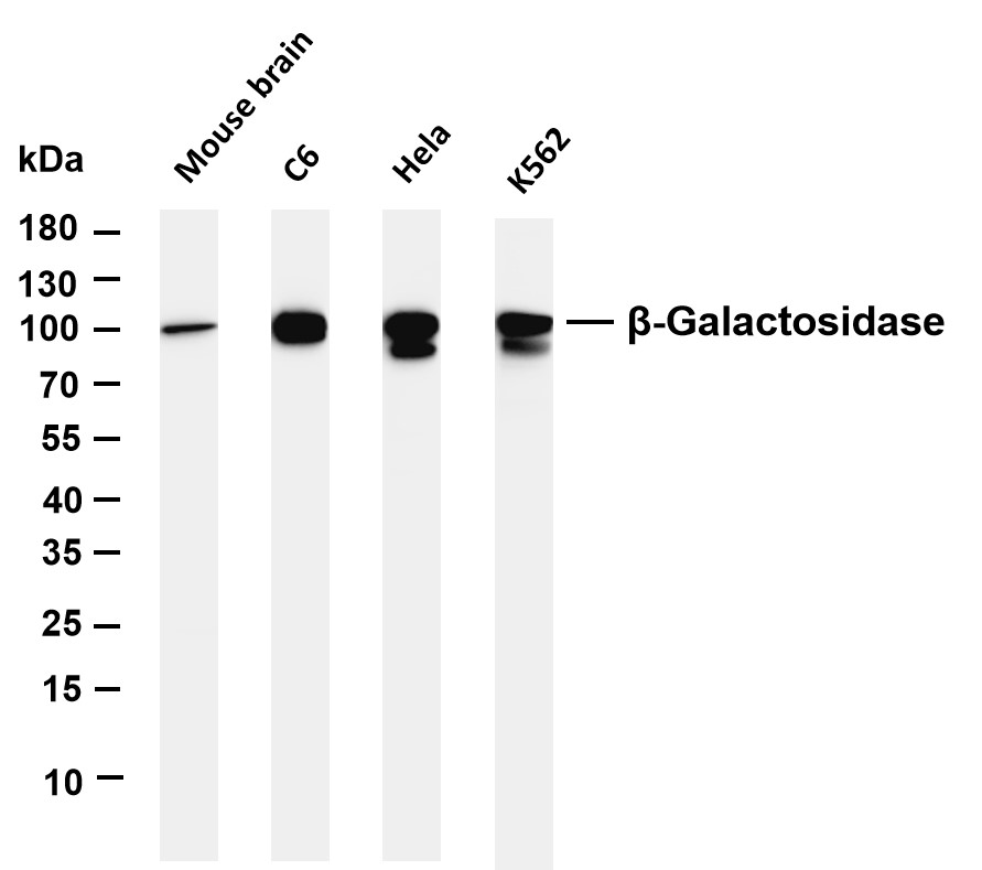
Various whole cell lysates were separated by 4-20% SDS-PAGE, and the membrane was blotted with anti-β-Galactosidase antibody. The HRP conjugated Goat anti-Rabbit IgG (H + L) antibody was used to detect the antibody.IHC
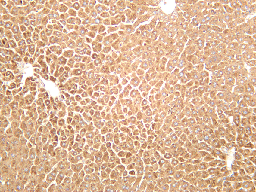
Mouse liver was stained with anti-β-Galactosidase rabbit antibody.IHC
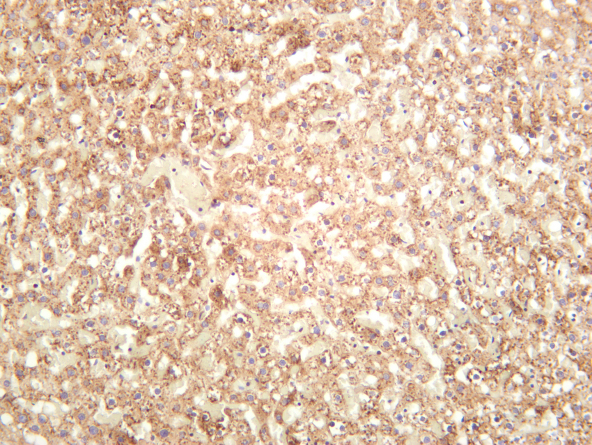
Rat liver was stained with anti-β-Galactosidase rabbit antibody.ICC/IF
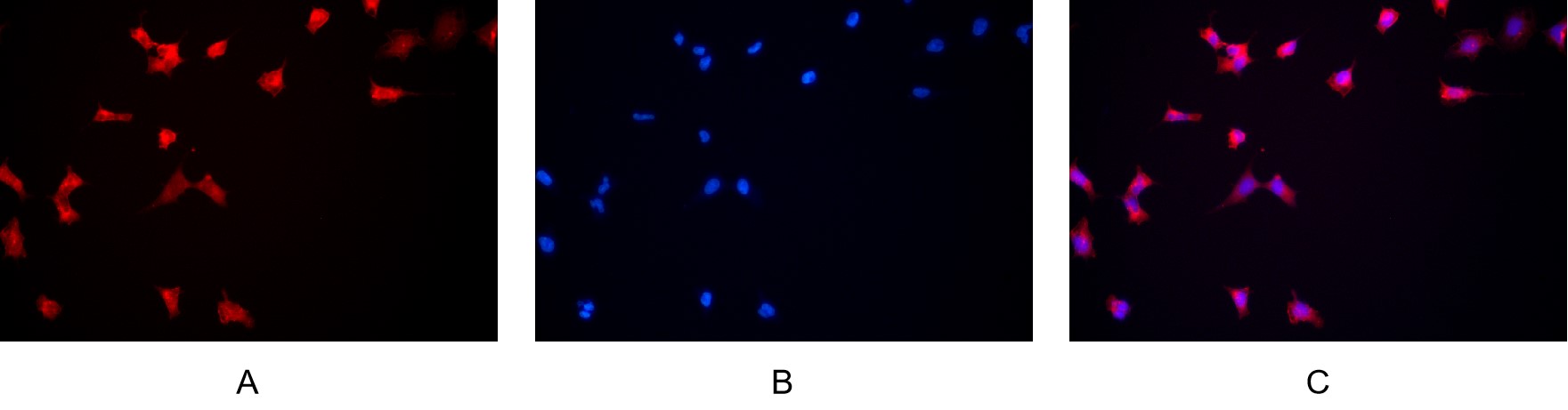
Immunofluorescence analysis of HEK293. Picture A: β-Galactosidase antibody (red). Picture B: DAPI (blue). Picture C: Merge of A+B .| Product Name | β-Galactosidase Rabbit mAb |
|---|---|
| Antibody Type | Primary Antibodies |
| Immunogen | The specific immunogen used to produce this antibody is proprietary information. |
| Clonality | Monoclonal |
|---|---|
| Isotype | IgG,K |
| Host Species | Rabbit |
| Tested Applications | ICC/IFIHCWB |
| WB:1:1000-1:5000 IHC:1:2000-1:10000 ICC/IF:1:200-1:1000 |
|
| Species Reactivity | HumanMouseRat |
| Concentration | 1mg/ml |
| Purification | Protein A |
| Gene Symbol | GLB1 |
|---|---|
| Gene Synonyms | EBP ELNR1 MPS4B |
| Gene Full Name | galactosidase beta 1 |
| Gene Summary | This gene encodes a member of the glycosyl hydrolase 35 family of proteins. Alternative splicing results in multiple transcript variants, at least one of which encodes a preproprotein that is proteolytically processed to generate the mature lysosomal enzyme. This enzyme catalyzes the hydrolysis of a terminal beta-linked galactose residue from ganglioside substrates and other glycoconjugates. Mutations in this gene may result in GM1-gangliosidosis and Morquio B syndrome. [provided by RefSeq, Nov 2015] |
| Alternative Names | GLB1 ELNR1 Beta-galactosidase Acid beta-galactosidase Lactase Elastin receptor 1 |
| Molecular Weight(MW) | 76 kDa(Observed band size: 100 kDa) |
| Cellular Localization | Lysosome |
WB
Various whole cell lysates were separated by 4-20% SDS-PAGE, and the membrane was blotted with anti-β-Galactosidase antibody. The HRP conjugated Goat anti-Rabbit IgG (H + L) antibody was used to detect the antibody.
IHC
Mouse liver was stained with anti-β-Galactosidase rabbit antibody.
IHC
Rat liver was stained with anti-β-Galactosidase rabbit antibody.
ICC/IF
Immunofluorescence analysis of HEK293. Picture A: β-Galactosidase antibody (red). Picture B: DAPI (blue). Picture C: Merge of A+B .| Application Notes | WB:1:1000-1:5000 IHC:1:2000-1:10000 ICC/IF:1:200-1:1000 |
|---|
| Form | Liquid |
|---|---|
| Storage Instructions | -15°C to -25°C/1 year (Do not lower than -25°C) |
| Storage Buffer | PBS, 50% glycerol, 0.05% Proclin 300, 0.05%BSA |


