WB
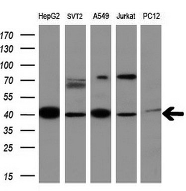
Western blot analysis of extracts (10ug) from 5 different cell lines by using anti-MMP13 monoclonal antibody at 1:200 dilution.WB
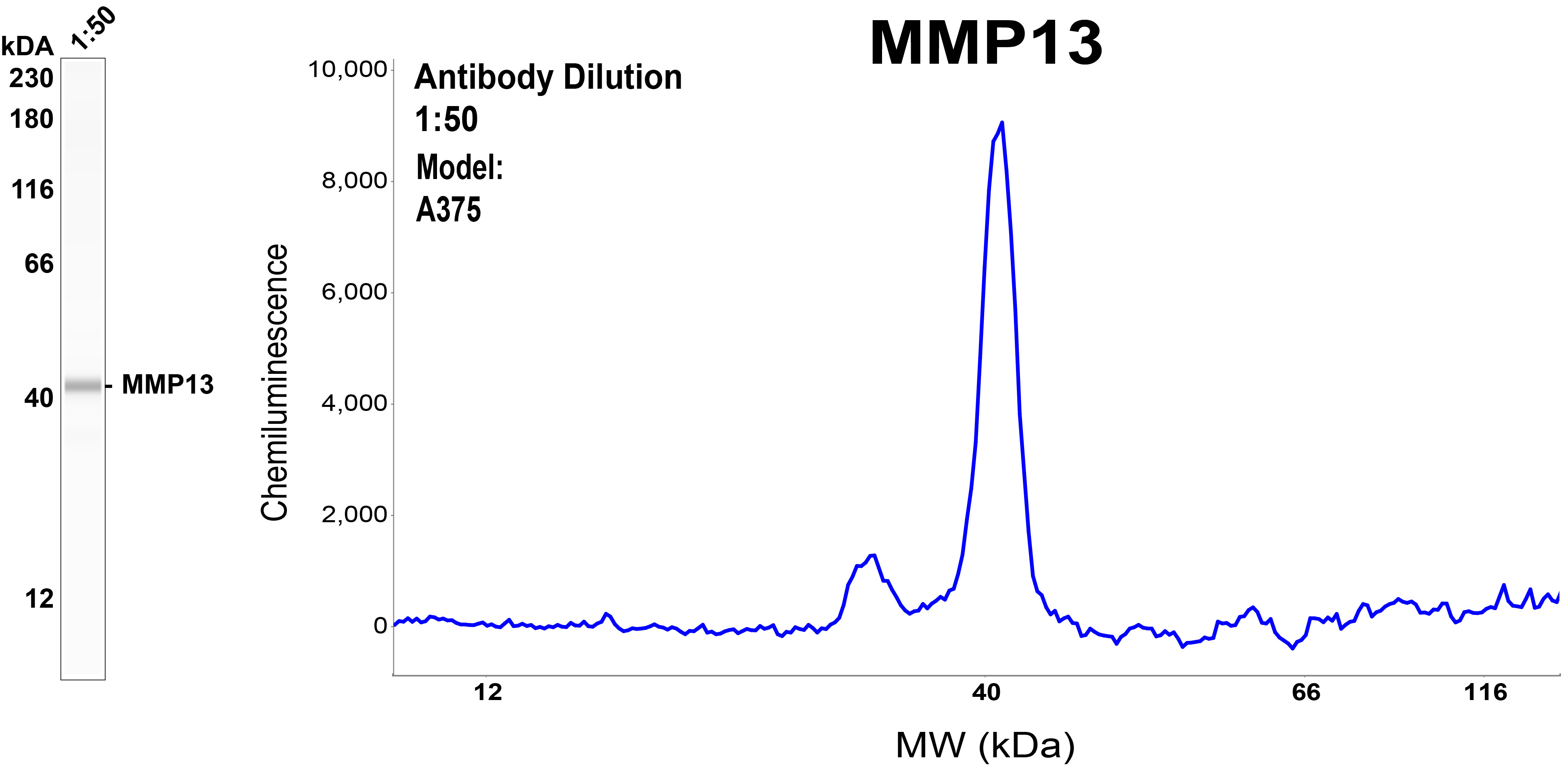
Simple Western™ analysis of endogenous protein MMP13 from A375 cell lysate (1 mg/mL) using MMP13 Mouse Monoclonal Antibody. The virtual lane view (left) shows the target (as indicated) at 1:50 dilution of primary antibody. The corresponding electropherogram view (right) plots chemiluminescence by molecular weight along the capillary at a 1:50 dilution of primary antibody. This experiment was performed under reducing conditions on the Simple Western instrument , using the Separation Module.IHC
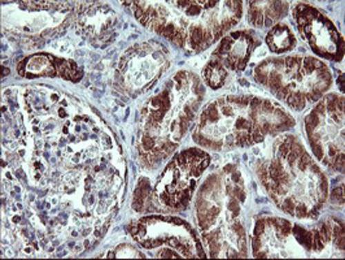
Immunohistochemical staining of paraffin-embedded Human Kidney tissue within the normal limits using anti-MMP13 mouse monoclonal antibody. Heat-induced epitope retrieval by EDTA solution buffer pH 8.0 at 120°C for 3 min.ICC/IF
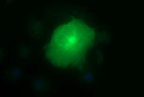
Anti-MMP13 mouse monoclonal antibody immunofluorescent staining of COS7 cells transiently transfected by pCMV6-ENTRY MMP13.| Product Name | MMP13 Mouse Monoclonal Antibody |
|---|---|
| Antibody Type | Primary Antibodies |
| Immunogen | Human recombinant protein fragment corresponding to amino acids 104-471 of human MMP13 (NP_002418) produced in HEK293T cell. |
| Clonality | monoclonal |
|---|---|
| Isotype | IgG2a |
| Host Species | Mouse |
| Tested Applications | ICC/IFIHCWB |
| WB:1:4000 IHC:1:150 ICC/IF:1:100 |
|
| Species Reactivity | HumanMouseRat |
| Concentration | 1mg/ml |
| Purification | Protein A/G |
| Gene Symbol | MMP13 |
|---|---|
| Gene Synonyms | CLG3 MDST MANDP1 MMP-13 |
| Gene Full Name | matrix metallopeptidase 13 |
| Gene Summary | This gene encodes a member of the peptidase M10 family of matrix metalloproteinases (MMPs). Proteins in this family are involved in the breakdown of extracellular matrix in normal physiological processes, such as embryonic development, reproduction, and tissue remodeling, as well as in disease processes, such as arthritis and metastasis. The encoded preproprotein is proteolytically processed to generate the mature protease. This protease cleaves type II collagen more efficiently than types I and III. It may be involved in articular cartilage turnover and cartilage pathophysiology associated with osteoarthritis. Mutations in this gene are associated with metaphyseal anadysplasia. This gene is part of a cluster of MMP genes on chromosome 11. [provided by RefSeq, Jan 2016] |
| Alternative Names | CLG3 MANDP1 MDST MMP-13 |
| Molecular Weight(MW) | 42.2 kDa |
| Cellular Localization | Extracellular matrix,Secreted. |
WB
Western blot analysis of extracts (10ug) from 5 different cell lines by using anti-MMP13 monoclonal antibody at 1:200 dilution.
WB
Simple Western™ analysis of endogenous protein MMP13 from A375 cell lysate (1 mg/mL) using MMP13 Mouse Monoclonal Antibody. The virtual lane view (left) shows the target (as indicated) at 1:50 dilution of primary antibody. The corresponding electropherogram view (right) plots chemiluminescence by molecular weight along the capillary at a 1:50 dilution of primary antibody. This experiment was performed under reducing conditions on the Simple Western instrument , using the Separation Module.
IHC
Immunohistochemical staining of paraffin-embedded Human Kidney tissue within the normal limits using anti-MMP13 mouse monoclonal antibody. Heat-induced epitope retrieval by EDTA solution buffer pH 8.0 at 120°C for 3 min.
ICC/IF
Anti-MMP13 mouse monoclonal antibody immunofluorescent staining of COS7 cells transiently transfected by pCMV6-ENTRY MMP13.| Application Notes | WB:1:4000 IHC:1:150 ICC/IF:1:100 |
|---|
| Form | Liquid |
|---|---|
| Storage Instructions | Store at -20°C as received. |
| Storage Buffer | PBS (pH 7.3) containing 1% BSA, 50% glycerol and 0.02% sodium azide. |


