WB
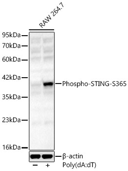
Western blot analysis of lysates from RAW 264.7 cells using Phospho-STING-S365 Rabbit pAb at 1:1000 dilution. RAW264.7 cells were transfection with poly(dA:dT) (5ug/mL) for 3 hours. Secondary antibody: HRP-conjugated Goat anti-Rabbit IgG (H+L) at 1:10000 dilution. Lysates/proteins: 25 μg per lane. Blocking buffer: 3% nonfat dry milk in TBST. Detection: ECL Basic Kit. Exposure time: 180s.ICC/IF
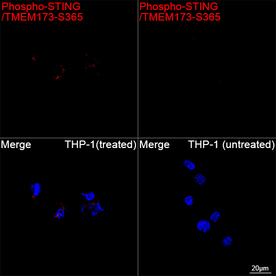
Confocal imaging of THP-1 cells (treated with PMA and poly(dA:dT)) and THP-1 cells (untreated) using Phospho-STING/TMEM173 S365 Rabbit pAb (dilution 1:1600) followed by a further incubation with Cy3 conjugated Goat anti-Rabbit IgG (H+L) (dilution 1:500) (Red). DAPI was used for nuclear staining (Blue). Objective: 100x.| Product Name | Phospho-STING/TMEM173-S365 Rabbit pAb |
|---|---|
| Antibody Type | Primary Antibodies |
| Immunogen | Synthetic peptide. This information is considered to be commercially sensitive. |
| Modification | p-S365 |
| Clonality | Polyclonal |
|---|---|
| Isotype | IgG |
| Host Species | Rabbit |
| Tested Applications | ICC/IFWB |
| WB:1:500 - 1:5000 IHC:1:1000 - 1:4000 ICC/IF:1:50-1:200 |
|
| Species Reactivity | HumanMouse |
| Concentration | 1mg/ml |
| Purification | Affinity purified |
| Gene Symbol | Sting1 |
|---|---|
| Gene Synonyms | ERIS MPYS Mita STING Tmem173 STING-beta 2610307O08Rik |
| Gene Full Name | stimulator of interferon response cGAMP interactor 1 |
| Gene Summary | Enables 2',3'-cyclic GMP-AMP binding activity; cyclic-di-GMP binding activity; and ubiquitin protein ligase binding activity. Involved in several processes, including defense response to other organism; macroautophagy; and positive regulation of innate immune response. Acts upstream of or within cellular response to interferon-beta; positive regulation of transcription by RNA polymerase II; and regulation of inflammatory response. Located in several cellular components, including Golgi membrane; autophagosome; and perinuclear region of cytoplasm. Part of STING complex. Is active in endoplasmic reticulum membrane and endoplasmic reticulum-Golgi intermediate compartment membrane. Is expressed in ductus deferens; epididymis; ileum; liver; and prostate gland. Used to study STING-associated vasculopathy with onset in infancy. Human ortholog(s) of this gene implicated in STING-associated vasculopathy with onset in infancy. Orthologous to human STING1 (stimulator of interferon response cGAMP interactor 1). [provided by Alliance of Genome Resources, Apr 2025] |
| Alternative Names | ERIS MPYS Mita STING Tmem173 STING-beta 2610307O08Rik Phospho-STING-S365 |
| Molecular Weight(MW) | 43 kDa(Observed band size: 41 kDa) |
| Cellular Localization | autophagosome, cytoplasm, cytosol, endoplasmic reticulum, endosome, Golgi apparatus, mitochondrial outer membrane, nucleoplasm, perinuclear region of cytoplasm, peroxisome, plasma membrane. |
WB
Western blot analysis of lysates from RAW 264.7 cells using Phospho-STING-S365 Rabbit pAb at 1:1000 dilution. RAW264.7 cells were transfection with poly(dA:dT) (5ug/mL) for 3 hours. Secondary antibody: HRP-conjugated Goat anti-Rabbit IgG (H+L) at 1:10000 dilution. Lysates/proteins: 25 μg per lane. Blocking buffer: 3% nonfat dry milk in TBST. Detection: ECL Basic Kit. Exposure time: 180s.
ICC/IF
Confocal imaging of THP-1 cells (treated with PMA and poly(dA:dT)) and THP-1 cells (untreated) using Phospho-STING/TMEM173 S365 Rabbit pAb (dilution 1:1600) followed by a further incubation with Cy3 conjugated Goat anti-Rabbit IgG (H+L) (dilution 1:500) (Red). DAPI was used for nuclear staining (Blue). Objective: 100x.| Application Notes | WB:1:500 - 1:5000 IHC:1:1000 - 1:4000 ICC/IF:1:50-1:200 |
|---|
| Form | Liquid |
|---|---|
| Storage Instructions | Store at -20℃. Avoid freeze / thaw cycles. |
| Storage Buffer | PBS, pH 7.3, containing 50% glycerol. Preserved with Proclin300 or sodium azide. May contain 0.05% BSA as specified on the Certificate of Analysis. |


