WB
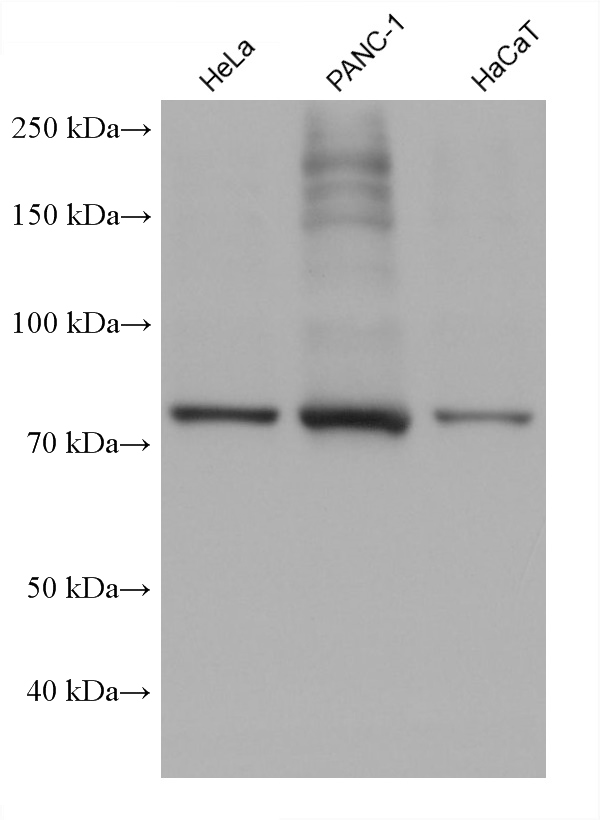
WB analysis using CPT1A Monoclonal antibody Various lysates were subjected to SDS PAGE followed by western blot with CPT1A Monoclonal antibody at dilution of 1:10000 incubated at room temperature for 1.5 hours.WB
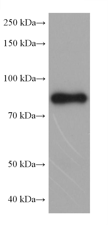
WB analysis of mouse liver using CPT1A Monoclonal antibody mouse liver tissue were subjected to SDS PAGE followed by western blot with CPT1A Monoclonal antibody at dilution of 1:10000 incubated at room temperature for 1.5 hours.IHC
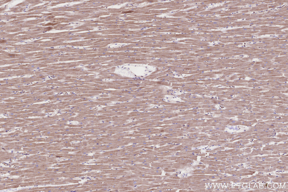
IHC staining of human heart using CPT1A Monoclonal antibody Immunohistochemical analysis of paraffin-embedded human heart tissue slide using CPT1A Monoclonal antibody at dilution of 1:2000 (under 10x lens). Heat mediated antigen retrieval with Tris-EDTA buffer (pH 9.0).IHC
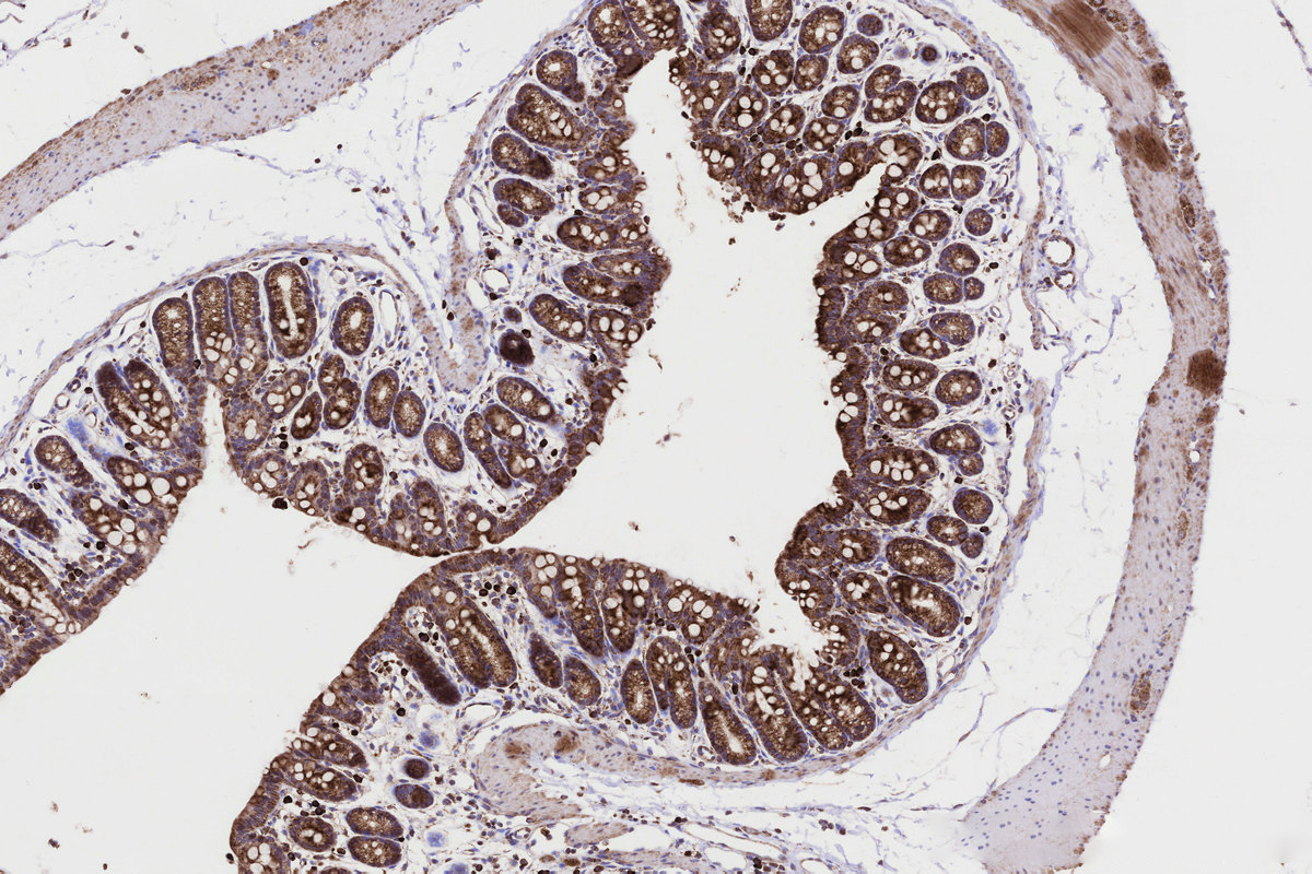
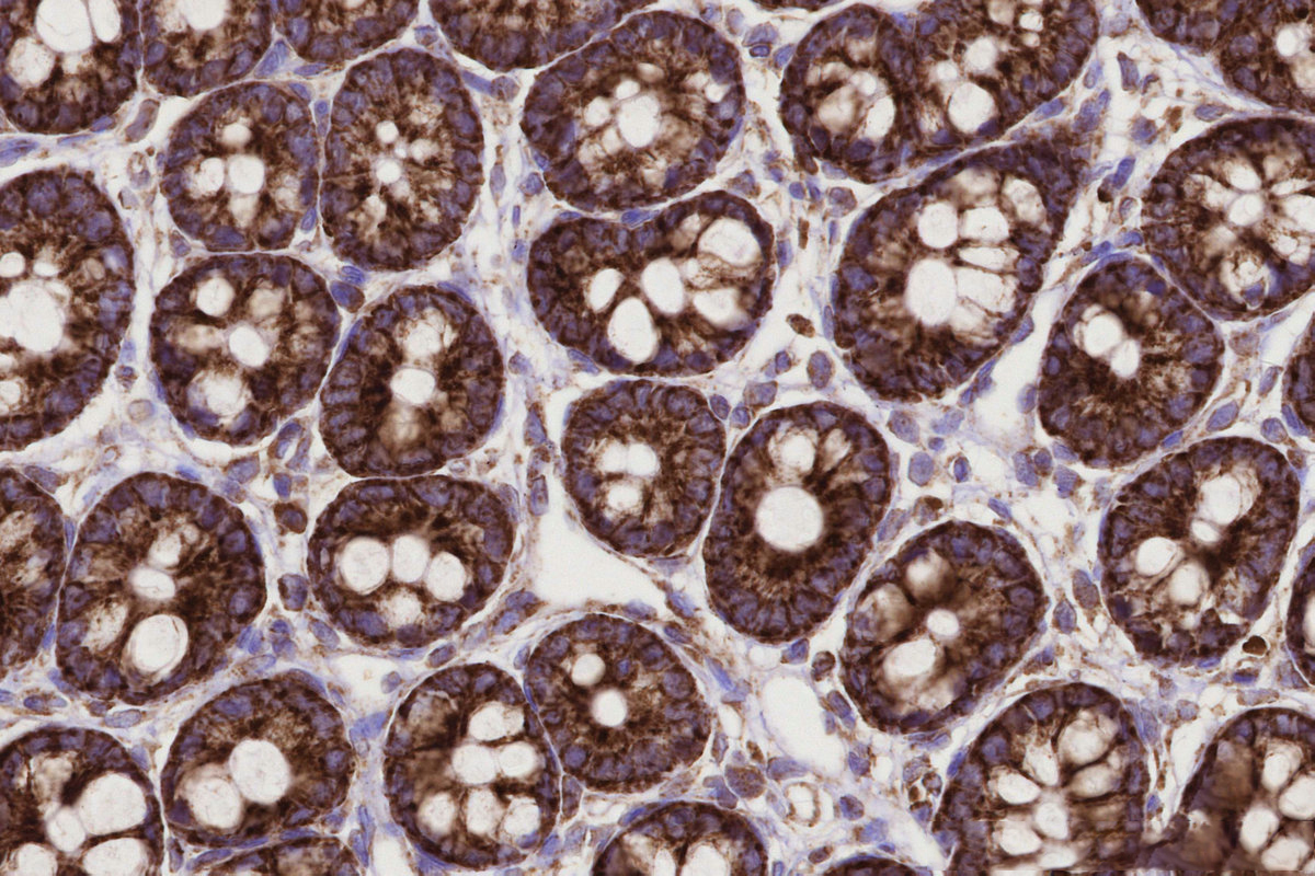
IHC staining of mouse colon using CPT1A Monoclonal antibody Immunohistochemical analysis of paraffin-embedded mouse colon tissue slide using CPT1A Monoclonal antibody at dilution of 1:2000 (under 10x lens). Heat mediated antigen retrieval with Tris-EDTA buffer (pH 9.0).IHC
IHC staining of rat colon using CPT1A Monoclonal antibody Immunohistochemical analysis of paraffin-embedded rat colon tissue slide using CPT1A Monoclonal antibody at dilution of 1:2000 (under 40x lens). Heat mediated antigen retrieval with Tris-EDTA buffer (pH 9.0).IP
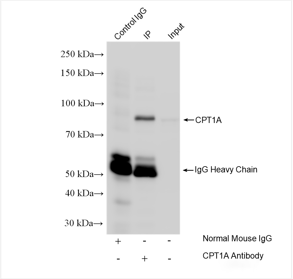
IP experiment of HepG2 using CPT1A Monoclonal antibody IP result of anti-CPT1A (4ug;1:3000) with HepG2 cells lysate 1800 ug.FC
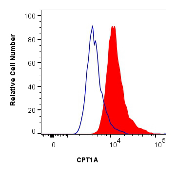
FC experiment of HeLa using CPT1A Monoclonal antibody 1x10^6 HeLa cells were intracellularly stained with 0.25 ug CPT1A Monoclonal antibody and 488-Conjugated Goat Anti-Mouse IgG(H+L) (red), or 0.25 ug Mouse IgG1 isotype control Mouse McAb (blue). Cells were fixed and permeabilized with Intracellular Flow Cytometry Fixation & Permeabilization Buffer Kit.| Product Name | CPT1A Monoclonal antibody |
|---|---|
| Antibody Type | Primary Antibodies |
| Immunogen | Recombinant human CPT1A protein |
| Clonality | Monoclonal |
|---|---|
| Isotype | IgG1 |
| Host Species | Mouse |
| Tested Applications | FCIHCIPWB |
| WB:1:5000-1:50000 IHC:1:500-1:2000 IP:0.5-4.0 ug for 1.0-3.0 mg of total protein lysate FC:0.25 ug per 10^6 cells in a 100 µl suspension |
|
| Species Reactivity | HumanMouseRat |
| Concentration | 1mg/ml |
| Purification | Protein G |
| Gene Symbol | CPT1A |
|---|---|
| Gene Synonyms | CPT1 CPT I CPT1-L CPTI-L L-CPT1 |
| Gene Full Name | carnitine palmitoyltransferase 1A |
| Gene Summary | The mitochondrial oxidation of long-chain fatty acids is initiated by the sequential action of carnitine palmitoyltransferase I (which is located in the outer membrane and is detergent-labile) and carnitine palmitoyltransferase II (which is located in the inner membrane and is detergent-stable), together with a carnitine-acylcarnitine translocase. CPT I is the key enzyme in the carnitine-dependent transport across the mitochondrial inner membrane and its deficiency results in a decreased rate of fatty acid beta-oxidation. Alternatively spliced transcript variants encoding different isoforms have been found for this gene. [provided by RefSeq, Jul 2008] |
| Alternative Names | 5D2B8 Carnitine O-palmitoyltransferase 1 liver isoform Carnitine O-palmitoyltransferase I liver isoform CPT I CPT1 |
| Molecular Weight(MW) | 88 kDa(Observed band size:86 kDa) |
| Cellular Localization | Membrane,Mitochondrion,Mitochondrion outer membrane. |
WB
WB analysis using CPT1A Monoclonal antibody Various lysates were subjected to SDS PAGE followed by western blot with CPT1A Monoclonal antibody at dilution of 1:10000 incubated at room temperature for 1.5 hours.
WB
WB analysis of mouse liver using CPT1A Monoclonal antibody mouse liver tissue were subjected to SDS PAGE followed by western blot with CPT1A Monoclonal antibody at dilution of 1:10000 incubated at room temperature for 1.5 hours.
IHC
IHC staining of human heart using CPT1A Monoclonal antibody Immunohistochemical analysis of paraffin-embedded human heart tissue slide using CPT1A Monoclonal antibody at dilution of 1:2000 (under 10x lens). Heat mediated antigen retrieval with Tris-EDTA buffer (pH 9.0).
IHC
IHC staining of mouse colon using CPT1A Monoclonal antibody Immunohistochemical analysis of paraffin-embedded mouse colon tissue slide using CPT1A Monoclonal antibody at dilution of 1:2000 (under 10x lens). Heat mediated antigen retrieval with Tris-EDTA buffer (pH 9.0).
IHC
IHC staining of rat colon using CPT1A Monoclonal antibody Immunohistochemical analysis of paraffin-embedded rat colon tissue slide using CPT1A Monoclonal antibody at dilution of 1:2000 (under 40x lens). Heat mediated antigen retrieval with Tris-EDTA buffer (pH 9.0).
IP
IP experiment of HepG2 using CPT1A Monoclonal antibody IP result of anti-CPT1A (4ug;1:3000) with HepG2 cells lysate 1800 ug.
FC
FC experiment of HeLa using CPT1A Monoclonal antibody 1x10^6 HeLa cells were intracellularly stained with 0.25 ug CPT1A Monoclonal antibody and 488-Conjugated Goat Anti-Mouse IgG(H+L) (red), or 0.25 ug Mouse IgG1 isotype control Mouse McAb (blue). Cells were fixed and permeabilized with Intracellular Flow Cytometry Fixation & Permeabilization Buffer Kit.| Application Notes | WB:1:5000-1:50000 IHC:1:500-1:2000 IP:0.5-4.0 ug for 1.0-3.0 mg of total protein lysate FC:0.25 ug per 10^6 cells in a 100 µl suspension |
|---|
| Form | Liquid |
|---|---|
| Storage Instructions | Store at -20°C. Stable for one year after shipment. Aliquoting is unnecessary for -20oC storage. |
| Storage Buffer | PBS with 0.02% sodium azide and 50% glycerol, pH 7.3. |


