WB
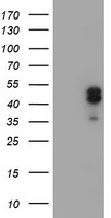
HEK293T cells were transfected with the pCMV6-ENTRY control , Left lane) or pCMV6-ENTRY LHX1, Right lane) cDNA for 48 hrs and lysed. Equivalent amounts of cell lysates (5 ug per lane) were separated by SDS-PAGE and immunoblotted with anti-LHX1.IHC
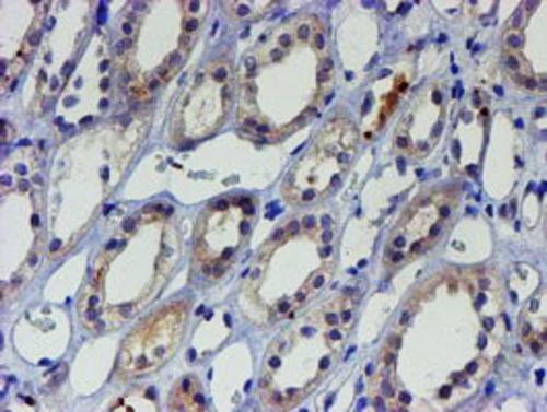
Immunohistochemical staining of paraffin embedded Human Kidney tissue within the normal limits using anti-LHX1 mouse monoclonal antibody. Heat-induced epitope retrieval by EDTA solution buffer pH 8.0 at 120°C for 3 min.ICC/IF
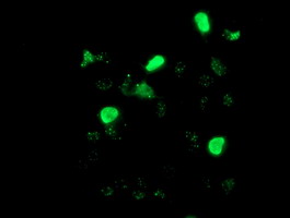
Anti-LHX1 mouse monoclonal antibody immunofluorescent staining of COS7 cells transiently transfected by pCMV6 ENTRY LHX1.FC
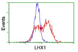
HEK293T cells transfected with either overexpress plasmid (Red) or empty vector control plasmid (Blue) were immunostained by anti-LHX1 antibody, and then analyzed by flow cytometry.| Product Name | LIM1 (LHX1) Mouse Monoclonal Antibody |
|---|---|
| Antibody Type | Primary Antibodies |
| Immunogen | Human recombinant protein fragment corresponding to amino acids 100-362 of human LHX1(NP_005559) produced in HEK293T cell. |
| Clonality | monoclonal |
|---|---|
| Isotype | IgG1 |
| Host Species | Mouse |
| Tested Applications | FCICC/IFIHCWB |
| WB:1:500-1:1000 IHC:1:150 ICC/IF:1:100 FC:1:100 |
|
| Species Reactivity | HumanMouseRat |
| Concentration | 1mg/ml |
| Purification | Protein A/G |
| Gene Symbol | LHX1 |
|---|---|
| Gene Synonyms | LIM1 LIM-1 |
| Gene Full Name | LIM homeobox 1 |
| Gene Summary | This gene encodes a member of a large protein family which contains the LIM domain, a unique cysteine-rich zinc-binding domain. The encoded protein is a transcription factor important for the development of the renal and urogenital systems. This gene is a candidate for Mayer-Rokitansky-Kuster-Hauser syndrome, a disorder characterized by anomalies in the female genital tract. [provided by RefSeq, Dec 2010] |
| Alternative Names | LIM-1 LIM1 |
| Molecular Weight(MW) | 44.6 kDa |
| Cellular Localization | Nucleus. |
WB
HEK293T cells were transfected with the pCMV6-ENTRY control , Left lane) or pCMV6-ENTRY LHX1, Right lane) cDNA for 48 hrs and lysed. Equivalent amounts of cell lysates (5 ug per lane) were separated by SDS-PAGE and immunoblotted with anti-LHX1.
IHC
Immunohistochemical staining of paraffin embedded Human Kidney tissue within the normal limits using anti-LHX1 mouse monoclonal antibody. Heat-induced epitope retrieval by EDTA solution buffer pH 8.0 at 120°C for 3 min.
ICC/IF
Anti-LHX1 mouse monoclonal antibody immunofluorescent staining of COS7 cells transiently transfected by pCMV6 ENTRY LHX1.
FC
HEK293T cells transfected with either overexpress plasmid (Red) or empty vector control plasmid (Blue) were immunostained by anti-LHX1 antibody, and then analyzed by flow cytometry.| Application Notes | WB:1:500-1:1000 IHC:1:150 ICC/IF:1:100 FC:1:100 |
|---|
| Form | Liquid |
|---|---|
| Storage Instructions | Store at -20°C as received. |
| Storage Buffer | PBS (pH 7.3) containing 1% BSA, 50% glycerol and 0.02% sodium azide. |


