WB
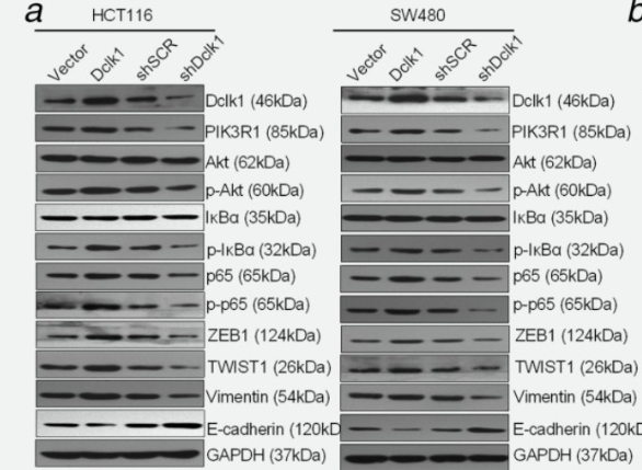
Liu, Weiying, et al. "DCLK1 promotes epithelial‐mesenchymal transition via the PI3K/Akt/NF‐κB pathway in colorectal cancer." International journal of cancer 142.10 (2018): 2068-2079.WB
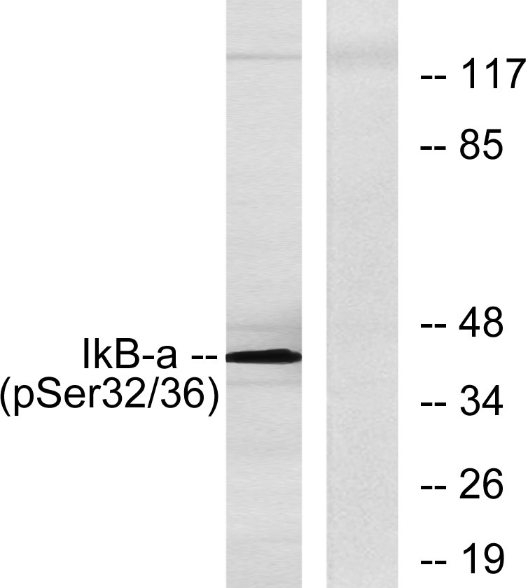
Western blot analysis of lysates from COS7 cells, using IkappaB-alpha (Phospho-Ser32/Ser36) Antibody. The lane on the right is blocked with the phospho peptide.IHC
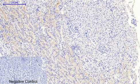
Immunohistochemical analysis of paraffin-embedded Rat-kidney tissue. 1,IκB α (phospho Ser32/S36) Polyclonal Antibody was diluted at 1:200 (4℃ overnight). 2, Sodium citrate pH 6.0 was used for antibody retrieval (>98℃,20min). 3,Secondary antibody was diluted at 1:200 (room tempeRature, 30min). Negative control was used by secondary antibody only.IHC
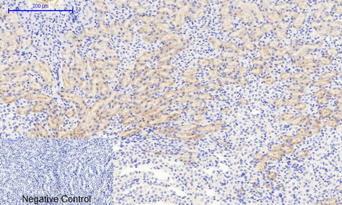
Immunohistochemical analysis of paraffin-embedded Mouse-kidney tissue. 1,IκB-α (phospho Ser32/S36) Polyclonal Antibody was diluted at 1:200 (4℃ overnight). 2, Sodium citrate pH 6.0 was used for antibody retrieval (>98℃,20min). 3,Secondary antibody was diluted at 1:200 (room tempeRature, 30min). Negative control was used by secondary antibody only.IHC
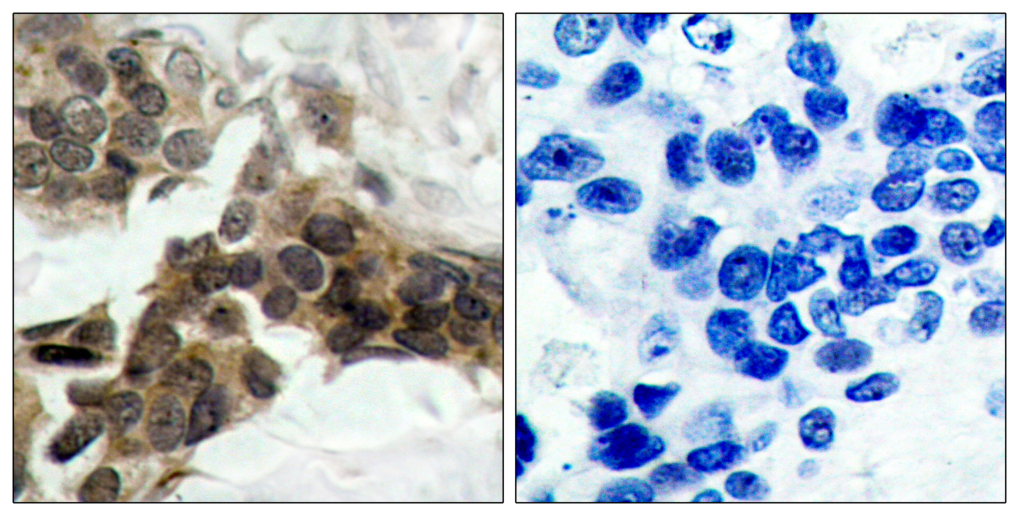
Immunohistochemistry analysis of paraffin-embedded human breast carcinoma, using IkappaB-alpha (Phospho-Ser32/Ser36) Antibody. The picture on the right is blocked with the phospho peptide.IF-P
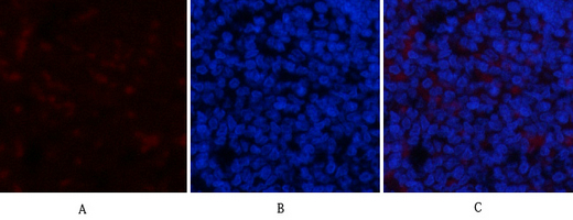
Immunofluorescence analysis of rat-spleen tissue. 1,IκB-α (phospho Ser32/S36) Polyclonal Antibody (red) was diluted at 1:200 (4℃ overnight). 2, Cy3 labled Secondary antibody was diluted at 1:300 (room temperature, 50min).3, Picture B: DAPI (blue) 10min. Picture A:Target. Picture B: DAPI. Picture C: merge of A+BICC/IF
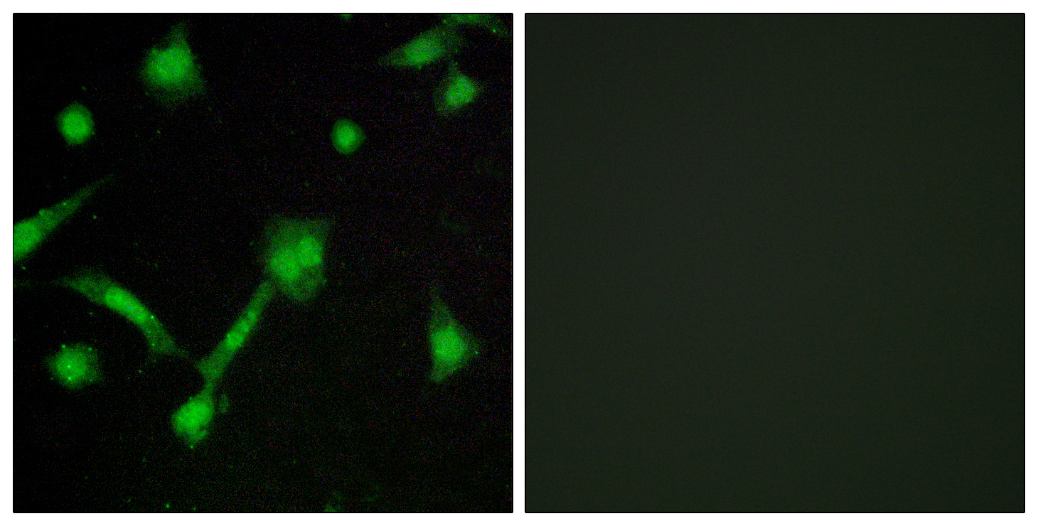
Immunofluorescence analysis of LOVO cells, using IkappaB-alpha (Phospho-Ser32/Ser36) Antibody. The picture on the right is blocked with the phospho peptide.| Product Name | IκB-α (Phospho Ser32/36) Rabbit pAb |
|---|---|
| Antibody Type | Primary Antibodies |
| Immunogen | The antiserum was produced against synthesized peptide derived from human IkappaB-alpha around the phosphorylation site of Ser32/Ser36. AA range:15-64 |
| Modification | p-Ser32/36 |
| Clonality | Polyclonal |
|---|---|
| Isotype | IgG |
| Host Species | Rabbit |
| Tested Applications | ICC/IFIF-PIHCWB |
| WB:1:500-1:2000 IHC:1:100-1:300 ICC/IF:1:200-1:1000 |
|
| Species Reactivity | HumanMonkeyMouseRat |
| Concentration | 1mg/ml |
| Purification | Affinity purified |
| Gene Symbol | NFKBIA |
|---|---|
| Gene Synonyms | IKBA MAD-3 NFKBI EDAID2 |
| Gene Full Name | NFKB inhibitor alpha |
| Gene Summary | This gene encodes a member of the NF-kappa-B inhibitor family, which contain multiple ankrin repeat domains. The encoded protein interacts with REL dimers to inhibit NF-kappa-B/REL complexes which are involved in inflammatory responses. The encoded protein moves between the cytoplasm and the nucleus via a nuclear localization signal and CRM1-mediated nuclear export. Mutations in this gene have been found in ectodermal dysplasia anhidrotic with T-cell immunodeficiency autosomal dominant disease. [provided by RefSeq, Aug 2011] |
| Alternative Names | NFKBIA IKBA MAD3 NFKBI NF-kappa-B inhibitor alpha I-kappa-B-alpha IkB-alpha IkappaBalpha Major histocompatibility complex enhancer-binding protein MAD3 |
| Molecular Weight(MW) | 40kDa |
| Cellular Localization | Cytoplasm. Nucleus. Shuttles between the nucleus and the cytoplasm by a nuclear localization signal (NLS) and a CRM1-dependent nuclear export. |
WB
Liu, Weiying, et al. "DCLK1 promotes epithelial‐mesenchymal transition via the PI3K/Akt/NF‐κB pathway in colorectal cancer." International journal of cancer 142.10 (2018): 2068-2079.
WB
Western blot analysis of lysates from COS7 cells, using IkappaB-alpha (Phospho-Ser32/Ser36) Antibody. The lane on the right is blocked with the phospho peptide.
IHC
Immunohistochemical analysis of paraffin-embedded Rat-kidney tissue. 1,IκB α (phospho Ser32/S36) Polyclonal Antibody was diluted at 1:200 (4℃ overnight). 2, Sodium citrate pH 6.0 was used for antibody retrieval (>98℃,20min). 3,Secondary antibody was diluted at 1:200 (room tempeRature, 30min). Negative control was used by secondary antibody only.
IHC
Immunohistochemical analysis of paraffin-embedded Mouse-kidney tissue. 1,IκB-α (phospho Ser32/S36) Polyclonal Antibody was diluted at 1:200 (4℃ overnight). 2, Sodium citrate pH 6.0 was used for antibody retrieval (>98℃,20min). 3,Secondary antibody was diluted at 1:200 (room tempeRature, 30min). Negative control was used by secondary antibody only.
IHC
Immunohistochemistry analysis of paraffin-embedded human breast carcinoma, using IkappaB-alpha (Phospho-Ser32/Ser36) Antibody. The picture on the right is blocked with the phospho peptide.
IF-P
Immunofluorescence analysis of rat-spleen tissue. 1,IκB-α (phospho Ser32/S36) Polyclonal Antibody (red) was diluted at 1:200 (4℃ overnight). 2, Cy3 labled Secondary antibody was diluted at 1:300 (room temperature, 50min).3, Picture B: DAPI (blue) 10min. Picture A:Target. Picture B: DAPI. Picture C: merge of A+B
ICC/IF
Immunofluorescence analysis of LOVO cells, using IkappaB-alpha (Phospho-Ser32/Ser36) Antibody. The picture on the right is blocked with the phospho peptide.| Application Notes | WB:1:500-1:2000 IHC:1:100-1:300 ICC/IF:1:200-1:1000 |
|---|
| Form | Liquid |
|---|---|
| Storage Instructions | -15°C to -25°C/1 year (Do not lower than -25°C) |
| Storage Buffer | Liquid in PBS containing 50% glycerol, 0.5% BSA and 0.02% sodium azide. |


