WB
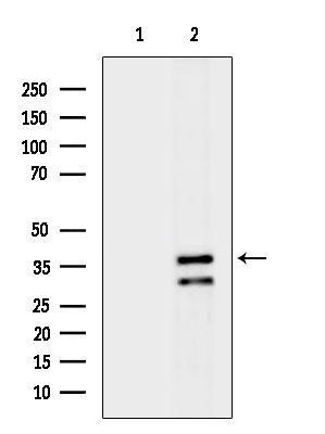
Western blot analysis of lysates from MC-3T3-E1 cells(H2O2 treatment), using SIAT4A Antibody. The lane on the left was treated with blocking peptide.IHC
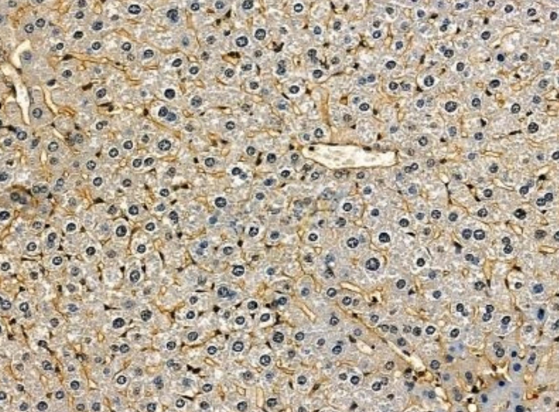
SIAT4A Antibody at 1/100 staining mouse liver tissue by IHC-P. The sample was formaldehyde fixed and a heat mediated antigen retrieval step in citrate buffer was performed. The sample was then blocked and incubated with the primary antibody at 4°C overnight. An HRP conjugated anti-rabbit antibody was used as the secondary antibody.ICC/IF
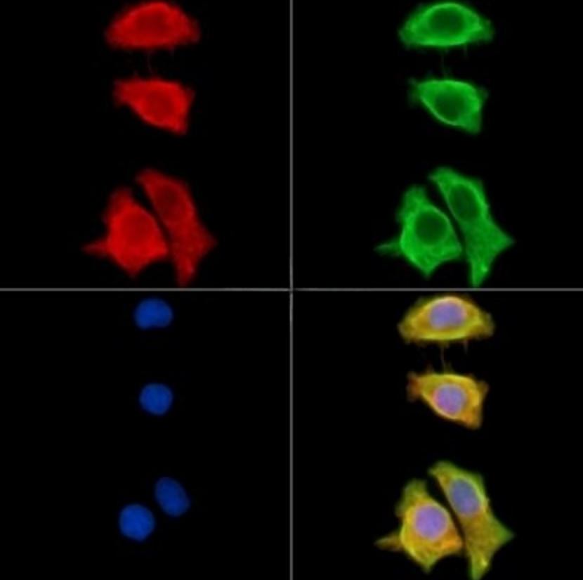
SIAT4A Antibody staining HepG2 cells by IF/ICC. The samples were fixed with PFA and permeabilized in 0.1% Triton X-100, then blocked in 10% serum for 45 minutes at 25°C. Samples were then incubated with primary Ab and mouse anti-beta tubulin Ab for 1 hour at 37°C. An AlexaFluor594 conjugated goat anti-rabbit IgG Ab(Red) and an AlexaFluor488 conjugated goat anti-mouse IgG Ab(Green) were used as the secondary antibody.The nuclear counter stain is DAPI (blue).| Product Name | Rabbit polyclonal antibody to SIAT4A |
|---|---|
| Antibody Type | Primary Antibodies |
| Immunogen | A synthesized peptide derived from Human SIAT4A. |
| Clonality | Polyclonal |
|---|---|
| Isotype | IgG |
| Host Species | Rabbit |
| Tested Applications | ICC/IFIHCWB |
| WB:1:500-1:2000 IHC:1:50-1:200 ICC/IF:1:100-1:500 |
|
| Species Reactivity | HumanMouse |
| Concentration | 1mg/ml |
| Purification | Affinity purified |
| Gene Symbol | ST3GAL1 |
|---|---|
| Gene Synonyms | ST3O SIAT4A SIATFL ST3GalA ST3GalIA Gal-NAc6S ST3GalA.1 ST3GalIA,1 |
| Gene Full Name | ST3 beta-galactoside alpha-2,3-sialyltransferase 1 |
| Gene Summary | The protein encoded by this gene is a type II membrane protein that catalyzes the transfer of sialic acid from CMP-sialic acid to galactose-containing substrates. The encoded protein is normally found in the Golgi but can be proteolytically processed to a soluble form. Correct glycosylation of the encoded protein may be critical to its sialyltransferase activity. This protein, which is a member of glycosyltransferase family 29, can use the same acceptor substrates as does sialyltransferase 4B. Two transcript variants encoding the same protein have been found for this gene. Other transcript variants may exist, but have not been fully characterized yet. [provided by RefSeq, Jul 2008] |
| Alternative Names | 3-GalNAc-alpha-2 3-sialyltransferase 1 3-sialyltransferase 3-ST 1 Alpha 2 Alpha 23 ST Beta galactoside alpha 23 sialyltransferase Beta-galactoside alpha-2 beta-galactoside alpha-2 3-sialyltransferase 1 CMP N acetylneuraminate beta galactosamide alpha 23 sialyltransferase CMP-N-acetylneuraminate-beta-galactosamide-alpha-2 DKFZp666E036 DKFZp779K2051 FLJ36548 Gal beta 13 GalNAc alpha 23 sialyltransferase Gal NAc6S Gal-beta-1 Gal-NAc6S MGC9183 OTTHUMP00000226912 OTTHUMP00000226913 SIA4A_HUMAN Sialyltransferase 4A SIAT4-A SIAT4A SIATFL ST3Gal I ST3GAL1 ST3GalA ST3GalA.1 ST3GalI ST3GalIA ST3O |
| Molecular Weight(MW) | 40 kDa |
| Cellular Localization | Golgi apparatus>Golgi stack membrane>Single-pass type II membrane protein. Secreted. Note: Membrane-bound form in trans cisternae of Golgi. Secreted into the body fluid. |
WB
Western blot analysis of lysates from MC-3T3-E1 cells(H2O2 treatment), using SIAT4A Antibody. The lane on the left was treated with blocking peptide.
IHC
SIAT4A Antibody at 1/100 staining mouse liver tissue by IHC-P. The sample was formaldehyde fixed and a heat mediated antigen retrieval step in citrate buffer was performed. The sample was then blocked and incubated with the primary antibody at 4°C overnight. An HRP conjugated anti-rabbit antibody was used as the secondary antibody.
ICC/IF
SIAT4A Antibody staining HepG2 cells by IF/ICC. The samples were fixed with PFA and permeabilized in 0.1% Triton X-100, then blocked in 10% serum for 45 minutes at 25°C. Samples were then incubated with primary Ab and mouse anti-beta tubulin Ab for 1 hour at 37°C. An AlexaFluor594 conjugated goat anti-rabbit IgG Ab(Red) and an AlexaFluor488 conjugated goat anti-mouse IgG Ab(Green) were used as the secondary antibody.The nuclear counter stain is DAPI (blue).| Application Notes | WB:1:500-1:2000 IHC:1:50-1:200 ICC/IF:1:100-1:500 |
|---|
| Form | Liquid |
|---|---|
| Storage Instructions | Store at -20 °C. Stable for 12 months from date of receipt. |
| Storage Buffer | Rabbit IgG in phosphate buffered saline , pH 7.4, 150mM NaCl, 0.02% sodium azide and 50% glycerol. |


