WB
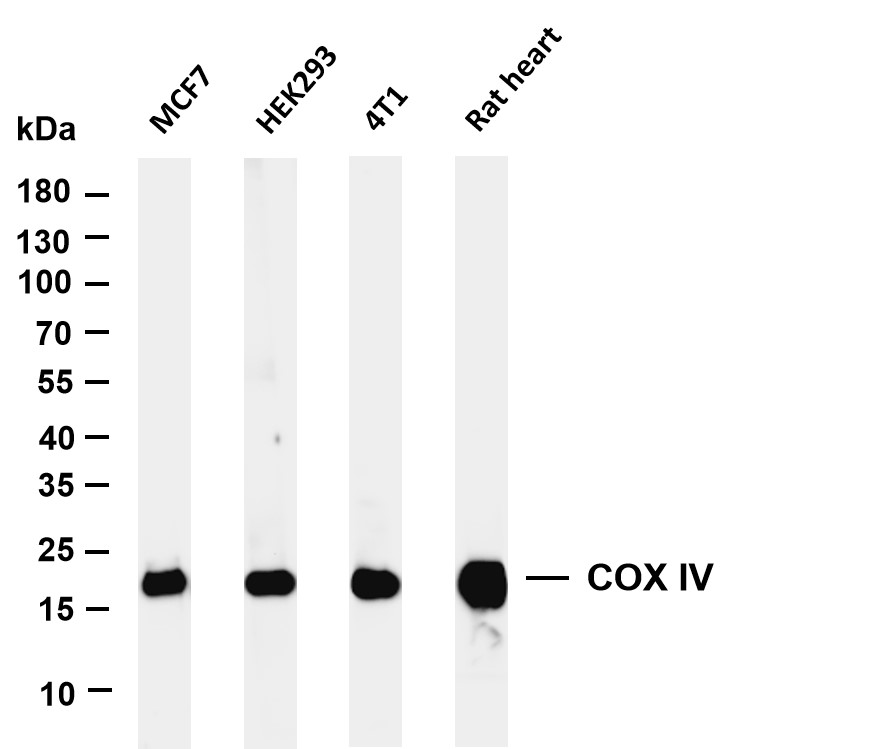
Various whole cell lysates were separated by 4-20% SDS-PAGE, and the membrane was blotted with anti-COX IV antibody. The HRP-conjugated Goat anti-Mouse IgG (H + L) antibody was used to detect the antibody.IHC
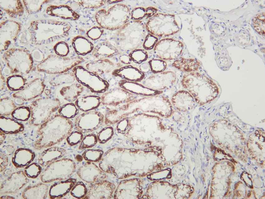
Human kidney was stained with anti-COX IV mouse antibodyIHC
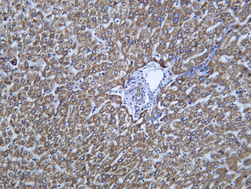
Human liver was stained with anti-COX IV Mouse antibodyICC/IF
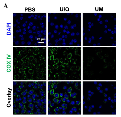
Immunogenic Radiation Therapy for Enhanced Antitumor Immunity via a Core–Shell Nanosensitizer-Mediated Immunosuppressive Tumor Microenvironment Modulation. ACS Nano Jin-Xiang Chen IF Mouse 4T1 cell-xenograftICC/IF
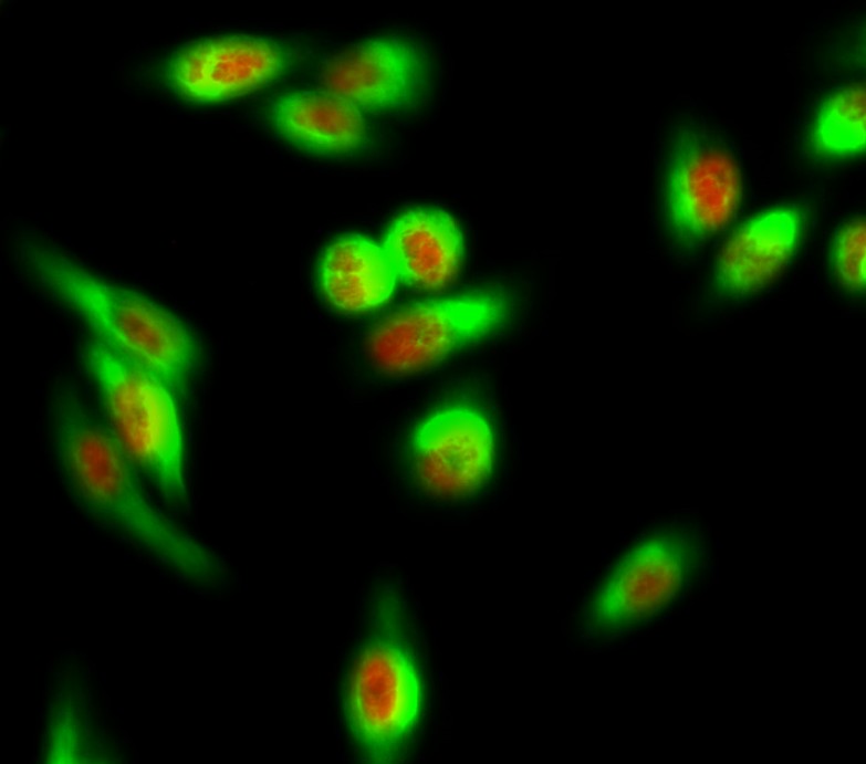
Immunofluorescence analysis of Hela cell. 1, Rabbit Hisitone H3 Antibody (red) was diluted at 1:200 (4℃ overnight).COX IV Mouse mAb (green) was diluted at 1:200 (4℃ overnight). 2, Goat Anti Rabbit Alexa Fluor 594 was diluted at 1:1000 (room temperature, 50min). Goat Anti Mouse Alexa Fluor 488 was diluted at 1:1000 (room temperature, 50min).IF-P
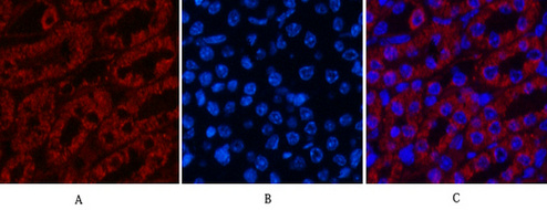
Immunofluorescence analysis of Mouse-kidney tissue. 1,COX IV Monoclonal Antibody (6C8) (red) was diluted at 1:200 (4℃ overnight). 2, Cy3 labled Secondary antibody was diluted at 1:300 (room temperature, 50min).3, Picture B: DAPI (blue) 10min. Picture A:Target. Picture B: DAPI. Picture C: merge of A+BIF-P
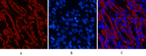
Immunofluorescence analysis of Rat-kidney tissue. 1,COX IV Monoclonal Antibody (6C8) (red) was diluted at 1:200 (4℃ overnight). 2, Cy3 labled Secondary antibody was diluted at 1:300 (room temperature, 50min).3, Picture B: DAPI (blue) 10min. Picture A:Target. Picture B: DAPI. Picture C: merge of A+B| Product Name | COX IV Mouse mAb |
|---|---|
| Antibody Type | Primary Antibodies |
| Immunogen | The specific immunogen used to produce this antibody is proprietary information. |
| Clonality | Monoclonal |
|---|---|
| Isotype | IgG2b,k |
| Host Species | Mouse |
| Tested Applications | ICC/IFIF-PIHCWB |
| WB:1:500-1:2000 IHC:1:200-1:1000 ICC/IF:1:100-500 IF-P:1:100-500 |
|
| Species Reactivity | HumanMouseRat |
| Concentration | 1mg/ml |
| Purification | Affinity purified |
| Gene Symbol | COX4I1 |
|---|---|
| Gene Synonyms | COX4 COXIV COX4-1 COXIV-1 MC4DN16 COX IV-1 |
| Gene Full Name | cytochrome c oxidase subunit 4I1 |
| Gene Summary | Cytochrome c oxidase (COX) is the terminal enzyme of the mitochondrial respiratory chain. It is a multi-subunit enzyme complex that couples the transfer of electrons from cytochrome c to molecular oxygen and contributes to a proton electrochemical gradient across the inner mitochondrial membrane. The complex consists of 13 mitochondrial- and nuclear-encoded subunits. The mitochondrially-encoded subunits perform the electron transfer and proton pumping activities. The functions of the nuclear-encoded subunits are unknown but they may play a role in the regulation and assembly of the complex. This gene encodes the nuclear-encoded subunit IV isoform 1 of the human mitochondrial respiratory chain enzyme. It is located at the 3' of the NOC4 (neighbor of COX4) gene in a head-to-head orientation, and shares a promoter with it. Pseudogenes related to this gene are located on chromosomes 13 and 14. Alternative splicing results in multiple transcript variants encoding different isoforms. [provided by RefSeq, Jan 2016] |
| Alternative Names | COX4I1 COX4 Cytochrome c oxidase subunit 4 isoform 1 mitochondrial Cytochrome c oxidase polypeptide IV Cytochrome c oxidase subunit IV isoform 1 COX IV-1 |
| Molecular Weight(MW) | 20 kDa(Observed band size: 17 kDa) |
| Cellular Localization | Nucleus. |
WB
Various whole cell lysates were separated by 4-20% SDS-PAGE, and the membrane was blotted with anti-COX IV antibody. The HRP-conjugated Goat anti-Mouse IgG (H + L) antibody was used to detect the antibody.
IHC
Human kidney was stained with anti-COX IV mouse antibody
IHC
Human liver was stained with anti-COX IV Mouse antibody
ICC/IF
Immunogenic Radiation Therapy for Enhanced Antitumor Immunity via a Core–Shell Nanosensitizer-Mediated Immunosuppressive Tumor Microenvironment Modulation. ACS Nano Jin-Xiang Chen IF Mouse 4T1 cell-xenograft
ICC/IF
Immunofluorescence analysis of Hela cell. 1, Rabbit Hisitone H3 Antibody (red) was diluted at 1:200 (4℃ overnight).COX IV Mouse mAb (green) was diluted at 1:200 (4℃ overnight). 2, Goat Anti Rabbit Alexa Fluor 594 was diluted at 1:1000 (room temperature, 50min). Goat Anti Mouse Alexa Fluor 488 was diluted at 1:1000 (room temperature, 50min).
IF-P
Immunofluorescence analysis of Mouse-kidney tissue. 1,COX IV Monoclonal Antibody (6C8) (red) was diluted at 1:200 (4℃ overnight). 2, Cy3 labled Secondary antibody was diluted at 1:300 (room temperature, 50min).3, Picture B: DAPI (blue) 10min. Picture A:Target. Picture B: DAPI. Picture C: merge of A+B
IF-P
Immunofluorescence analysis of Rat-kidney tissue. 1,COX IV Monoclonal Antibody (6C8) (red) was diluted at 1:200 (4℃ overnight). 2, Cy3 labled Secondary antibody was diluted at 1:300 (room temperature, 50min).3, Picture B: DAPI (blue) 10min. Picture A:Target. Picture B: DAPI. Picture C: merge of A+B| Application Notes | WB:1:500-1:2000 IHC:1:200-1:1000 ICC/IF:1:100-500 IF-P:1:100-500 |
|---|
| Form | Liquid |
|---|---|
| Storage Instructions | -15°C to -25°C/1 year(Do not lower than -25°C) |
| Storage Buffer | PBS, 50% glycerol, 0.05% Proclin 300, 0.05%BSA |


