WB
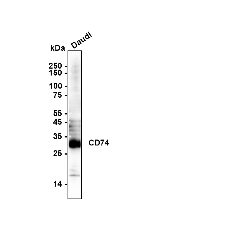
Western blot analysis of CD74 on different lysates with Mouse anti-CD74 antibody at 1/1,000 dilution. Lysates/proteins at 20 µg/Lane. Exposure time: 29 Seconds; Blocking: 5% NFDM/TBST, 1 hour at room temperature. Primary antibody: 1/1,000 in primary antibody dilution buffer, overnight at 4 ℃. Secondary antibody: Goat anti-Mouse IgG-HRP, 1/50,000 in 5% NFDM/TBST, 1 hour at room temperature.IHC
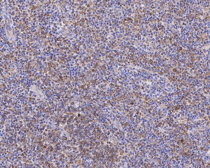
Immunohistochemical analysis of paraffin-embedded human B-cell lymphoma tissue with Mouse anti-CD74 antibody at 1/5,000 dilution. The section was pre-treated using heat mediated antigen retrieval with Tris-EDTA buffer (pH 9.0) for 20 minutes. The tissues were blocked in 1% BSA for 20 minutes at room temperature, washed with ddH2O and PBS, and then probed with the primary antibody at 1/5,000 dilution for 1 hour at room temperature. The detection was performed using an HRP conjugated compact polymer system. DAB was used as the chromogen. Tissues were counterstained with hematoxylin and mounted with DPX.ICC/IF
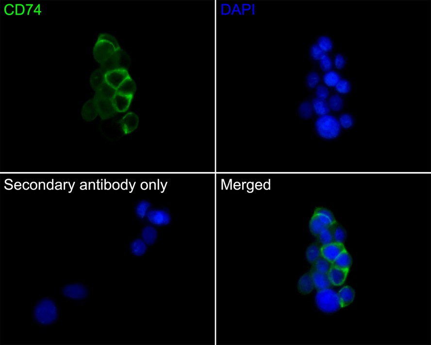
Immunocytochemistry analysis of LOVO cells labeling CD74 with Mouse anti-CD74 antibody at 1/50 dilution. Cells were fixed in 4% paraformaldehyde for 30 minutes, permeabilized with 0.1% Triton X-100 in PBS for 15 minutes, and then blocked with 2% BSA for 30 minutes at room temperature. Cells were then incubated with Mouse anti-CD74 antibody at 1/50 dilution in 2% BSA overnight at 4 ℃. Goat Anti-Mouse IgG H&L was used as the secondary antibody at 1/1,000 dilution. PBS instead of the primary antibody was used as the secondary antibody only control. Nuclear DNA was labelled in blue with DAPI.IF-P
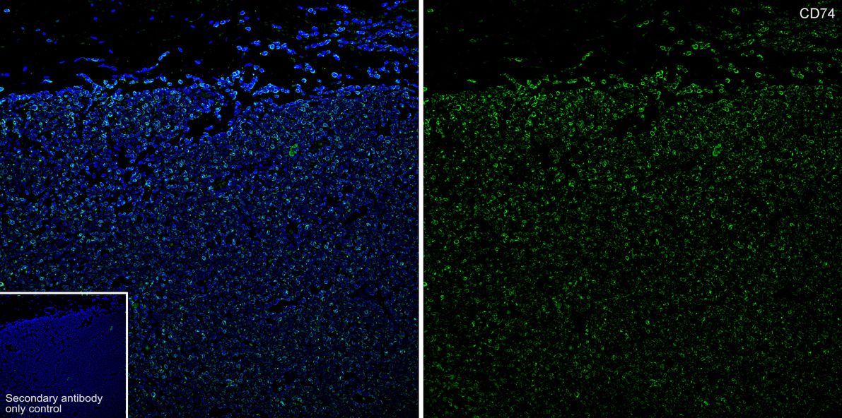
Application: IF-Tissue. Species: Human. Site: B-cell lymphoma. Sample: Paraffin-embedded section. Antibody concentration: 1/1,000.| Product Name | CD74 Recombinant Mouse Monoclonal Antibody |
|---|---|
| Antibody Type | Primary Antibodies |
| Immunogen | Recombinant protein within Human CD74 73-296 (Extracellular). |
| Clonality | Monoclonal |
|---|---|
| Isotype | IgG1 |
| Host Species | Mouse |
| Tested Applications | ICC/IFIF-PIHCWB |
| WB:1:1000 IHC:1:1000 ICC/IF:1:50 IF-P:1:1000 |
|
| Species Reactivity | Human |
| Concentration | 1mg/ml |
| Purification | Protein A |
| Gene Symbol | CD74 |
|---|---|
| Gene Synonyms | II p33 CLIP DHLAG HLADG Ia-GAMMA |
| Gene Full Name | CD74 molecule |
| Gene Summary | The protein encoded by this gene associates with class II major histocompatibility complex (MHC) and is an important chaperone that regulates antigen presentation for immune response. It also serves as cell surface receptor for the cytokine macrophage migration inhibitory factor (MIF) which, when bound to the encoded protein, initiates survival pathways and cell proliferation. This protein also interacts with amyloid precursor protein (APP) and suppresses the production of amyloid beta (Abeta). Multiple alternatively spliced transcript variants encoding different isoforms have been identified. [provided by RefSeq, Aug 2011] |
| Alternative Names | CD 74 antibody CD74 antibody CD74 antigen (invariant polypeptide of major histocompatibility complex class II antigen-associated) antibody CD74 antigen antibody CD74 molecule antibody CD74 molecule major histocompatibility complex class II invariant chain antibody CLIP antibody DHLAG antibody Gamma chain of class II antigens antibody HG2A_HUMAN antibody HLA class II histocompatibility antigen gamma chain antibody HLA DR antigens associated invariant chain antibody HLA DR gamma antibody HLA-DR antigens-associated invariant chain antibody HLA-DR-gamma antibody HLADG antibody HLADR antigens associated invariant chain antibody Ia antigen associated invariant chain antibody Ia antigen-associated invariant chain antibody Ia associated invariant chain antibody Ia gamma antibody Ii antibody Invariant polypeptide of major histocompatibility complex class II antigen associated antibody la-gamma antibody Major histocompatibility complex class II invariant chain antibody MHC HLA DR gamma chain antibody MHC HLA-DR gamma chain antibody p33 antibody p35 antibody Protein 41 antibody |
| Molecular Weight(MW) | 33.5 kDa |
| Cellular Localization | Endoplasmic reticulum membrane, trans-Golgi network, cell membrane, lysosome, endosome. |
WB
Western blot analysis of CD74 on different lysates with Mouse anti-CD74 antibody at 1/1,000 dilution. Lysates/proteins at 20 µg/Lane. Exposure time: 29 Seconds; Blocking: 5% NFDM/TBST, 1 hour at room temperature. Primary antibody: 1/1,000 in primary antibody dilution buffer, overnight at 4 ℃. Secondary antibody: Goat anti-Mouse IgG-HRP, 1/50,000 in 5% NFDM/TBST, 1 hour at room temperature.
IHC
Immunohistochemical analysis of paraffin-embedded human B-cell lymphoma tissue with Mouse anti-CD74 antibody at 1/5,000 dilution. The section was pre-treated using heat mediated antigen retrieval with Tris-EDTA buffer (pH 9.0) for 20 minutes. The tissues were blocked in 1% BSA for 20 minutes at room temperature, washed with ddH2O and PBS, and then probed with the primary antibody at 1/5,000 dilution for 1 hour at room temperature. The detection was performed using an HRP conjugated compact polymer system. DAB was used as the chromogen. Tissues were counterstained with hematoxylin and mounted with DPX.
ICC/IF
Immunocytochemistry analysis of LOVO cells labeling CD74 with Mouse anti-CD74 antibody at 1/50 dilution. Cells were fixed in 4% paraformaldehyde for 30 minutes, permeabilized with 0.1% Triton X-100 in PBS for 15 minutes, and then blocked with 2% BSA for 30 minutes at room temperature. Cells were then incubated with Mouse anti-CD74 antibody at 1/50 dilution in 2% BSA overnight at 4 ℃. Goat Anti-Mouse IgG H&L was used as the secondary antibody at 1/1,000 dilution. PBS instead of the primary antibody was used as the secondary antibody only control. Nuclear DNA was labelled in blue with DAPI.
IF-P
Application: IF-Tissue. Species: Human. Site: B-cell lymphoma. Sample: Paraffin-embedded section. Antibody concentration: 1/1,000.| Application Notes | WB:1:1000 IHC:1:1000 ICC/IF:1:50 IF-P:1:1000 |
|---|
| Form | Liquid |
|---|---|
| Storage Instructions | Shipped at 4℃. Store at +4℃ short term (1-2 weeks). Store at -20℃ long term. |
| Storage Buffer | PBS (pH7.4), 0.1% BSA, 40% Glycerol. Preservative: 0.05% Sodium Azide. |


