WB
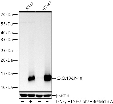
Western blot analysis of various lysates using CXCL10/IP-10 Rabbit mAb at 1:10000 dilution incubated overnight at 4℃. A549 cells and HT-29 cells were treated with TNF-α (20ng/mL) for 6 hours, Brefeldin A (300ng/mL) for 3 hours and IFN-γ (50ng/mL) for 8 hours. Secondary antibody: HRP-conjugated Goat anti-Rabbit IgG (H+L) at 1:10000 dilution. Lysates/proteins: 30 μg per lane. Blocking buffer: 3% nonfat dry milk in TBST. Detection: ECL Basic Kit. Exposure time: 5s.IHC
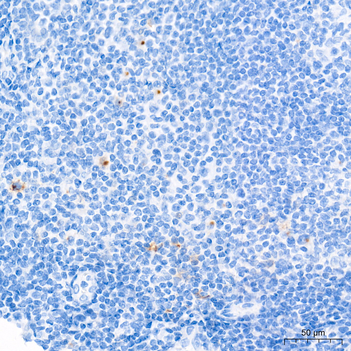
Immunohistochemistry analysis of paraffin embedded Human tonsil tissue using CXCL10/IP-10 Rabbit mAb at a dilution of 1:500 (40x lens). High pressure antigen retrieval performed with 0.01M Tris EDTA Buffer (pH 9.0) prior to IHC staining.ICC/IF
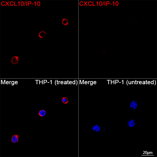
Confocal imaging of THP-1 cells (treated with IFN-γ, LPS and BFA) and THP-1 cells (untreated) using CXCL10/IP-10 Rabbit mAb (dilution 1:200) followed by a further incubation with Cy3 Goat Anti-Rabbit IgG (H+L) (dilution 1:500) (Red). DAPI was used for nuclear staining (Blue). Objective: 100x.| Product Name | CXCL10/IP-10 Rabbit mAb |
|---|---|
| Antibody Type | Primary Antibodies |
| Immunogen | Recombinant protein (or fragment).This information is considered to be commercially sensitive. |
| Clonality | Monoclonal |
|---|---|
| Isotype | IgG |
| Host Species | Rabbit |
| Tested Applications | ICC/IFIHCWB |
| WB:1:10000 - 1:60000 IHC:1:200 - 1:800 ICC/IF:1:100 - 1:400 |
|
| Species Reactivity | Human |
| Concentration | 1mg/ml |
| Purification | Affinity purified |
| Gene Symbol | CXCL10 |
|---|---|
| Gene Synonyms | C7 IFI10 INP10 IP-10 crg-2 mob-1 SCYB10 gIP-10 |
| Gene Full Name | C-X-C motif chemokine ligand 10 |
| Gene Summary | This antimicrobial gene encodes a chemokine of the CXC subfamily and ligand for the receptor CXCR3. Binding of this protein to CXCR3 results in pleiotropic effects, including stimulation of monocytes, natural killer and T-cell migration, and modulation of adhesion molecule expression. This gene may also be a key regulator of the 'cytokine storm' immune response to SARS-CoV-2 infection. [provided by RefSeq, Sep 2020] |
| Alternative Names | C7 IFI10 INP10 IP-10 crg-2 mob-1 SCYB10 gIP-10 CXCL10/IP-10 |
| Molecular Weight(MW) | 11 kDa |
| Cellular Localization | Secreted. |
WB
Western blot analysis of various lysates using CXCL10/IP-10 Rabbit mAb at 1:10000 dilution incubated overnight at 4℃. A549 cells and HT-29 cells were treated with TNF-α (20ng/mL) for 6 hours, Brefeldin A (300ng/mL) for 3 hours and IFN-γ (50ng/mL) for 8 hours. Secondary antibody: HRP-conjugated Goat anti-Rabbit IgG (H+L) at 1:10000 dilution. Lysates/proteins: 30 μg per lane. Blocking buffer: 3% nonfat dry milk in TBST. Detection: ECL Basic Kit. Exposure time: 5s.
IHC
Immunohistochemistry analysis of paraffin embedded Human tonsil tissue using CXCL10/IP-10 Rabbit mAb at a dilution of 1:500 (40x lens). High pressure antigen retrieval performed with 0.01M Tris EDTA Buffer (pH 9.0) prior to IHC staining.
ICC/IF
Confocal imaging of THP-1 cells (treated with IFN-γ, LPS and BFA) and THP-1 cells (untreated) using CXCL10/IP-10 Rabbit mAb (dilution 1:200) followed by a further incubation with Cy3 Goat Anti-Rabbit IgG (H+L) (dilution 1:500) (Red). DAPI was used for nuclear staining (Blue). Objective: 100x.| Application Notes | WB:1:10000 - 1:60000 IHC:1:200 - 1:800 ICC/IF:1:100 - 1:400 |
|---|
| Form | Liquid |
|---|---|
| Storage Instructions | Store at -20℃. Avoid freeze / thaw cycles. |
| Storage Buffer | PBS containing 50% glycerol and 0.05% BSA, preserved with proclin300 or sodium azide (as specified on the Certificate of Analysis), pH 7.3. |


