WB
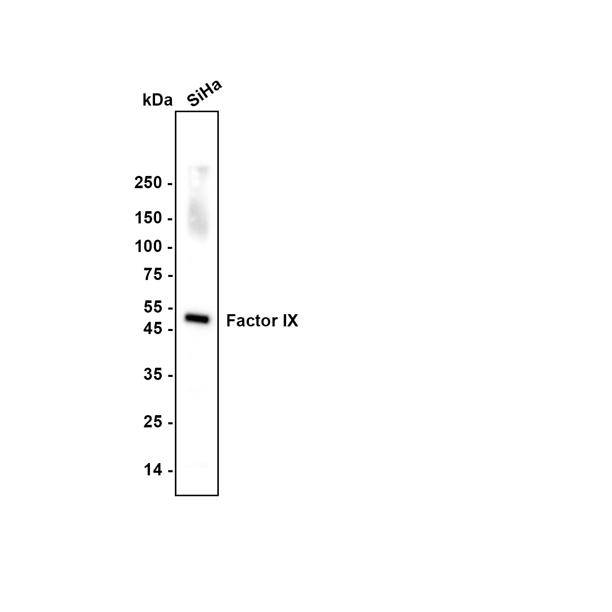
Western blot analysis of Factor IX on SiHa cell lysate with Rabbit anti-Factor IX antibody at 1/1,000 dilution. Lysates/proteins at 15 µg/Lane. Exposure time: 10 seconds; Blocking: 5% NFDM/TBST, 1 hour at room temperature Primary antibody: 1/1,000 in primary antibody dilution buffer, overnight at 4 ℃ Secondary antibody: Goat anti-Rabbit IgG-HRP, 1/50,000 in 5% NFDM/TBST, 1 hour at room temperatureIHC
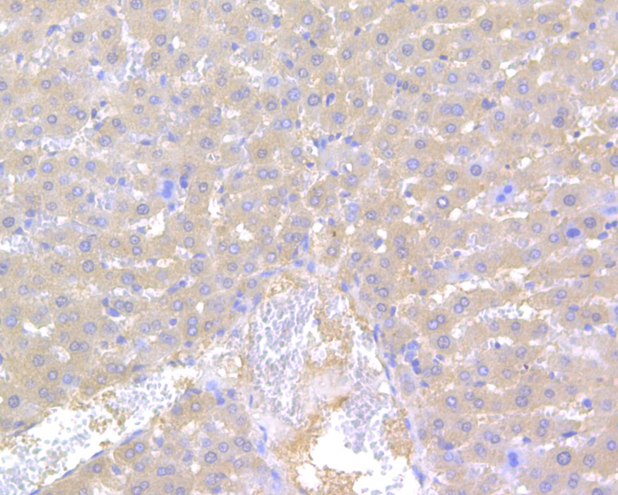
Immunohistochemical analysis of paraffin-embedded rat liver tissue using anti-Factor IX antibody. The section was pre-treated using heat mediated antigen retrieval with Tris-EDTA buffer (pH 8.0-8.4) for 20 minutes.The tissues were blocked in 5% BSA for 30 minutes at room temperature, washed with ddH2O and PBS, and then probed with the primary antibody (1/200) for 30 minutes at room temperature. The detection was performed using an HRP conjugated compact polymer system. DAB was used as the chromogen. Tissues were counterstained with hematoxylin and mounted with DPX.IHC
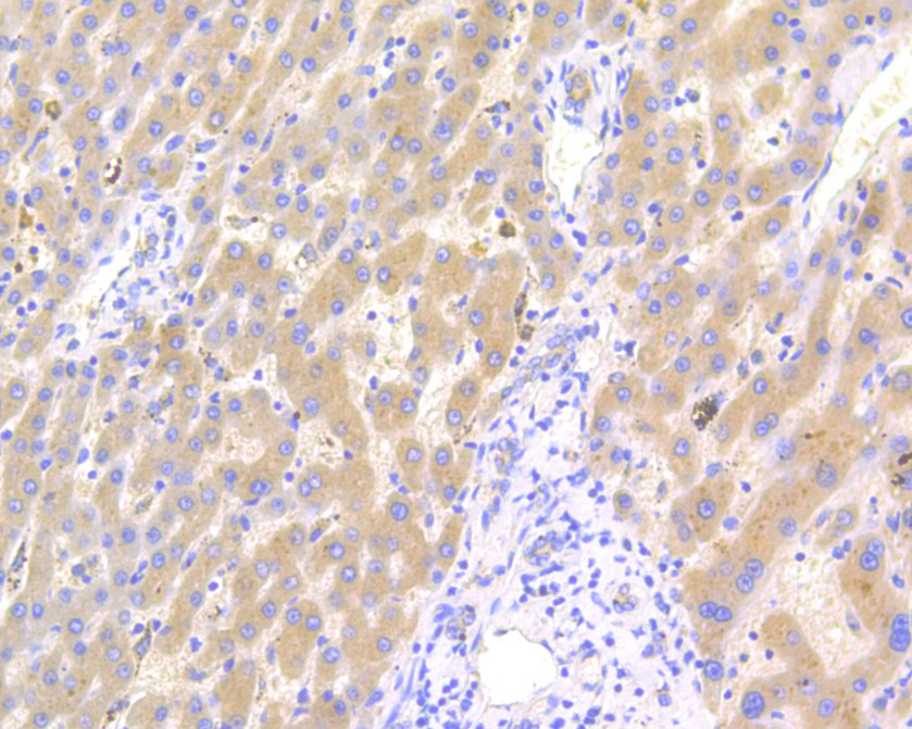
Immunohistochemical analysis of paraffin-embedded human liver tissue using anti-Factor IX antibody. The section was pre-treated using heat mediated antigen retrieval with Tris-EDTA buffer (pH 8.0-8.4) for 20 minutes.The tissues were blocked in 5% BSA for 30 minutes at room temperature, washed with ddH2O and PBS, and then probed with the primary antibody (1/800) for 30 minutes at room temperature. The detection was performed using an HRP conjugated compact polymer system. DAB was used as the chromogen. Tissues were counterstained with hematoxylin and mounted with DPX.IHC
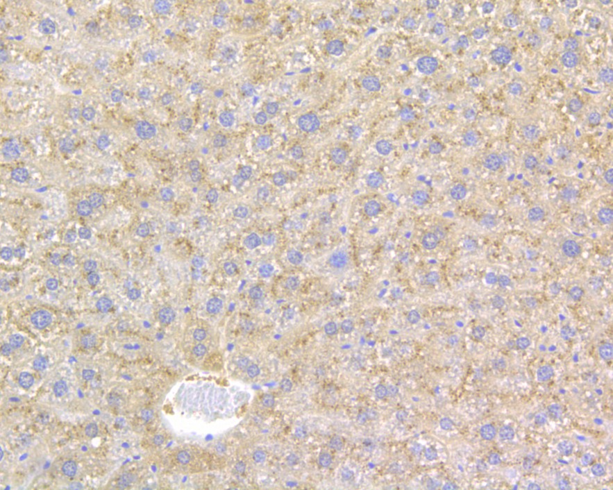
Immunohistochemical analysis of paraffin-embedded mouse liver tissue using anti-Factor IX antibody. The section was pre-treated using heat mediated antigen retrieval with Tris-EDTA buffer (pH 8.0-8.4) for 20 minutes.The tissues were blocked in 5% BSA for 30 minutes at room temperature, washed with ddH2O and PBS, and then probed with the primary antibody (1/800) for 30 minutes at room temperature. The detection was performed using an HRP conjugated compact polymer system. DAB was used as the chromogen. Tissues were counterstained with hematoxylin and mounted with DPX.FC
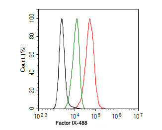
Flow cytometric analysis of SiHa cells labeling Factor IX. Cells were fixed and permeabilized. Then stained with the primary antibody (1ug/ml) (red) compared with Rabbit IgG Isotype Control (green). After incubation of the primary antibody at +4℃ for an hour, the cells were stained with a 488 conjugate-Goat anti-Rabbit IgG Secondary antibody at 1/1,000 dilution for 30 minutes at +4℃. Unlabelled sample was used as a control (cells without incubation with primary antibody; black).| Product Name | Factor IX Rabbit Polyclonal Antibody |
|---|---|
| Antibody Type | Primary Antibodies |
| Immunogen | Synthetic peptide within human Factor IX aa 20-70. |
| Clonality | Polyclonal |
|---|---|
| Isotype | IgG |
| Host Species | Rabbit |
| Tested Applications | FCIHCWB |
| WB:1:1000 IHC:1:100-1:500 FC:1:500-1:1000 |
|
| Species Reactivity | HumanMouseRat |
| Concentration | 1mg/ml |
| Purification | Affinity purified |
| Gene Symbol | F9 |
|---|---|
| Gene Synonyms | FIX P19 PTC HEMB THPH8 F9 p22 |
| Gene Full Name | coagulation factor IX |
| Gene Summary | This gene encodes vitamin K-dependent coagulation factor IX that circulates in the blood as an inactive zymogen. This factor is converted to an active form by factor XIa, which excises the activation peptide and thus generates a heavy chain and a light chain held together by one or more disulfide bonds. The role of this activated factor IX in the blood coagulation cascade is to activate factor X to its active form through interactions with Ca+2 ions, membrane phospholipids, and factor VIII. Alterations of this gene, including point mutations, insertions and deletions, cause factor IX deficiency, which is a recessive X-linked disorder, also called hemophilia B or Christmas disease. Alternative splicing results in multiple transcript variants encoding different isoforms that may undergo similar proteolytic processing. [provided by RefSeq, Sep 2015] |
| Alternative Names | Christmas Disease antibody Christmas factor antibody Coagulant factor IX antibody Coagulation factor 9 antibody Coagulation factor IX antibody Coagulation factor IXa heavy chain antibody F9 antibody FA9_HUMAN antibody Factor 9 antibody Factor IX Deficiency antibody FactorIX antibody FIX antibody Haemophilia B antibody HEMB antibody MGC129641 antibody MGC129642 antibody P19 antibody Plasma Thromboplastic Component antibody Plasma thromboplastin component antibody PTC antibody |
| Molecular Weight(MW) | 52 kDa |
| Cellular Localization | Secreted. |
WB
Western blot analysis of Factor IX on SiHa cell lysate with Rabbit anti-Factor IX antibody at 1/1,000 dilution. Lysates/proteins at 15 µg/Lane. Exposure time: 10 seconds; Blocking: 5% NFDM/TBST, 1 hour at room temperature Primary antibody: 1/1,000 in primary antibody dilution buffer, overnight at 4 ℃ Secondary antibody: Goat anti-Rabbit IgG-HRP, 1/50,000 in 5% NFDM/TBST, 1 hour at room temperature
IHC
Immunohistochemical analysis of paraffin-embedded rat liver tissue using anti-Factor IX antibody. The section was pre-treated using heat mediated antigen retrieval with Tris-EDTA buffer (pH 8.0-8.4) for 20 minutes.The tissues were blocked in 5% BSA for 30 minutes at room temperature, washed with ddH2O and PBS, and then probed with the primary antibody (1/200) for 30 minutes at room temperature. The detection was performed using an HRP conjugated compact polymer system. DAB was used as the chromogen. Tissues were counterstained with hematoxylin and mounted with DPX.
IHC
Immunohistochemical analysis of paraffin-embedded human liver tissue using anti-Factor IX antibody. The section was pre-treated using heat mediated antigen retrieval with Tris-EDTA buffer (pH 8.0-8.4) for 20 minutes.The tissues were blocked in 5% BSA for 30 minutes at room temperature, washed with ddH2O and PBS, and then probed with the primary antibody (1/800) for 30 minutes at room temperature. The detection was performed using an HRP conjugated compact polymer system. DAB was used as the chromogen. Tissues were counterstained with hematoxylin and mounted with DPX.
IHC
Immunohistochemical analysis of paraffin-embedded mouse liver tissue using anti-Factor IX antibody. The section was pre-treated using heat mediated antigen retrieval with Tris-EDTA buffer (pH 8.0-8.4) for 20 minutes.The tissues were blocked in 5% BSA for 30 minutes at room temperature, washed with ddH2O and PBS, and then probed with the primary antibody (1/800) for 30 minutes at room temperature. The detection was performed using an HRP conjugated compact polymer system. DAB was used as the chromogen. Tissues were counterstained with hematoxylin and mounted with DPX.
FC
Flow cytometric analysis of SiHa cells labeling Factor IX. Cells were fixed and permeabilized. Then stained with the primary antibody (1ug/ml) (red) compared with Rabbit IgG Isotype Control (green). After incubation of the primary antibody at +4℃ for an hour, the cells were stained with a 488 conjugate-Goat anti-Rabbit IgG Secondary antibody at 1/1,000 dilution for 30 minutes at +4℃. Unlabelled sample was used as a control (cells without incubation with primary antibody; black).| Application Notes | WB:1:1000 IHC:1:100-1:500 FC:1:500-1:1000 |
|---|
| Form | Liquid |
|---|---|
| Storage Instructions | Shipped at 4℃. Store at +4℃ short term (1-2 weeks). It is recommended to aliquot into single-use upon delivery. Store at -20℃ long term. |
| Storage Buffer | 1*TBS (pH7.4), 0.2% BSA, 50% Glycerol. Preservative: 0.05% Sodium Azide. |


