WB
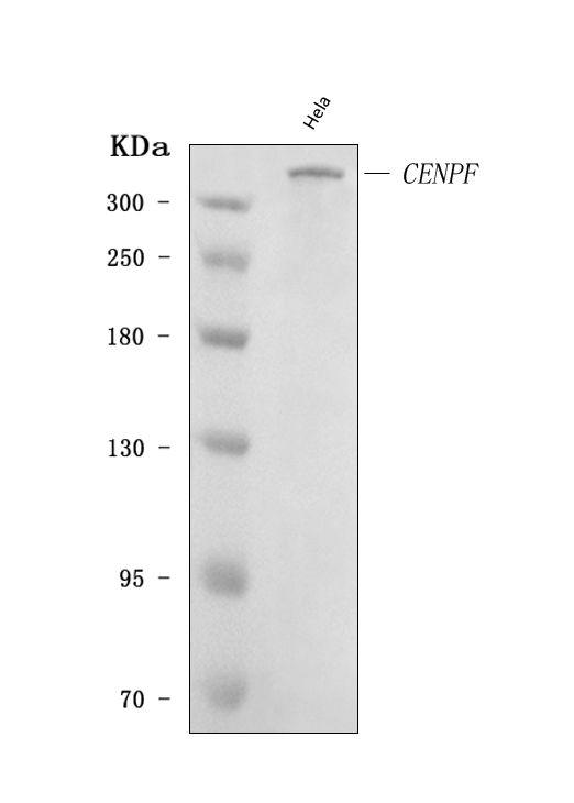
Western blot analysis of CENPF using anti-CENPF antibody. The sample well of each lane was loaded with 30 ug of sample under reducing conditions. After electrophoresis, proteins were transferred to a membrane. Then the membrane was incubated with rabbit anti-CENPF antigen affinity purified polyclonal antibody at a dilution of 1:1000 and probed with a goat anti-rabbit IgG-HRP secondary antibody. The signal is developed using ECL Plus Western Blotting Substrate.ICC/IF
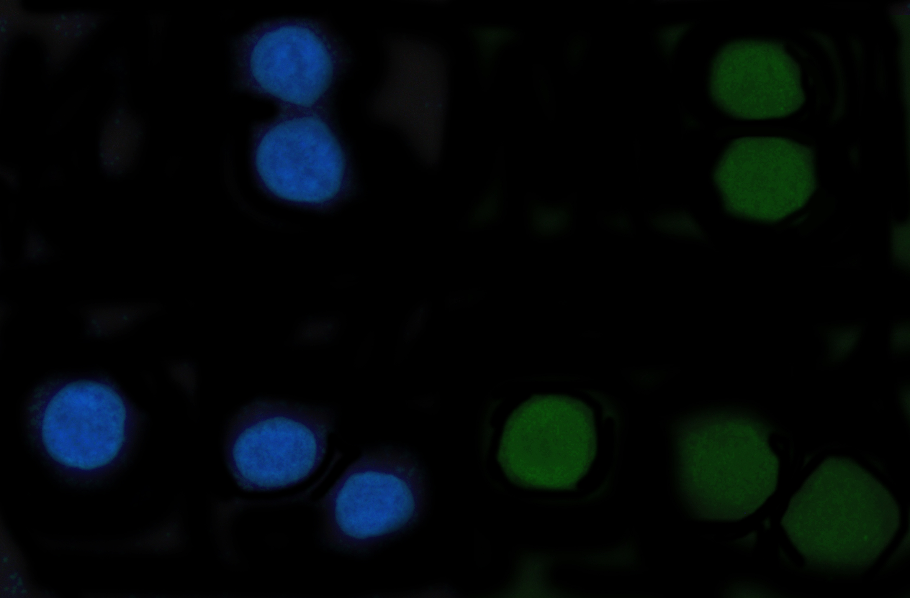
ICC/IF analysis of CENPF using anti-CENPF antibody. CENPF was detected in an immunocytochemical section of SiHa cells. The section was incubated with rabbit anti-CENPF Antibody at a dilution of 1:100. Fluoro488 Conjugated Goat Anti-Rabbit IgG (Green) was used as secondary antibody. The section was counterstained with DAPI (Blue).FC
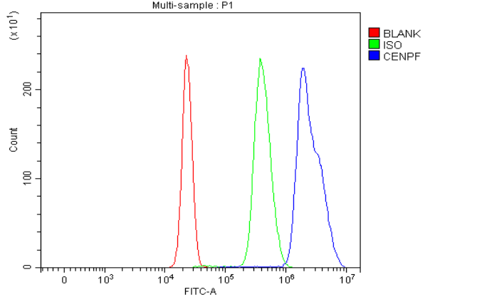
Flow Cytometry analysis of HepG2 cells using anti-CENPF antibody. Overlay histogram showing HepG2 cells stained with Rabbit polyclonal antibody to CENPF (Blue line). To facilitate intracellular staining, cells were fixed with 4% paraformaldehyde and permeabilized with permeabilization buffer. The cells were blocked with 10% normal goat serum. And then incubated with rabbit anti-CENPF Antibody at 1:100 dilution for 30 min at 20°C. Fluoro488 conjugated goat anti-rabbit IgG was used as secondary antibody at 1:100 dilution for 30 minutes at 20°C. Isotype control antibody (Green line) was rabbit IgG at 1:100 dilution used under the same conditions. Unlabelled sample without incubation with primary antibody and secondary antibody (Red line) was used as a blank control.| Product Name | Rabbit polyclonal antibody to CENPF |
|---|---|
| Antibody Type | Primary Antibodies |
| Immunogen | E.coli-derived human CENPF recombinant protein (Position: Q129-R3078). |
| Clonality | Polyclonal |
|---|---|
| Isotype | IgG |
| Host Species | Rabbit |
| Tested Applications | FCICC/IFWB |
| WB:1:500-1:2000 ICC/IF:1:50-1:400 FC:1:50-1:200 |
|
| Species Reactivity | Human |
| Concentration | 0.5mg/ml |
| Purification | Affinity purified |
| Gene Symbol | CENPF |
|---|---|
| Gene Synonyms | CENF hcp-1 CILD31 STROMS PRO1779 |
| Gene Full Name | centromere protein F |
| Gene Summary | This gene encodes a protein that associates with the centromere-kinetochore complex. The protein is a component of the nuclear matrix during the G2 phase of interphase. In late G2 the protein associates with the kinetochore and maintains this association through early anaphase. It localizes to the spindle midzone and the intracellular bridge in late anaphase and telophase, respectively, and is thought to be subsequently degraded. The localization of this protein suggests that it may play a role in chromosome segregation during mitotis. It is thought to form either a homodimer or heterodimer. Autoantibodies against this protein have been found in patients with cancer or graft versus host disease. [provided by RefSeq, Jul 2008] |
| Alternative Names | AH antigen CENF CENP F CENPF Centromere protein F hcp 1 Kinetochore protein CENPF Mitosin PRO1779 |
| Molecular Weight(MW) | 368 kDa(Observed band size:358 kDa) |
| Cellular Localization | Centromere,Chromosome,Cytoplasm,Cytoskeleton,Kinetochore,Nucleus. |
WB
Western blot analysis of CENPF using anti-CENPF antibody. The sample well of each lane was loaded with 30 ug of sample under reducing conditions. After electrophoresis, proteins were transferred to a membrane. Then the membrane was incubated with rabbit anti-CENPF antigen affinity purified polyclonal antibody at a dilution of 1:1000 and probed with a goat anti-rabbit IgG-HRP secondary antibody. The signal is developed using ECL Plus Western Blotting Substrate.
ICC/IF
ICC/IF analysis of CENPF using anti-CENPF antibody. CENPF was detected in an immunocytochemical section of SiHa cells. The section was incubated with rabbit anti-CENPF Antibody at a dilution of 1:100. Fluoro488 Conjugated Goat Anti-Rabbit IgG (Green) was used as secondary antibody. The section was counterstained with DAPI (Blue).
FC
Flow Cytometry analysis of HepG2 cells using anti-CENPF antibody. Overlay histogram showing HepG2 cells stained with Rabbit polyclonal antibody to CENPF (Blue line). To facilitate intracellular staining, cells were fixed with 4% paraformaldehyde and permeabilized with permeabilization buffer. The cells were blocked with 10% normal goat serum. And then incubated with rabbit anti-CENPF Antibody at 1:100 dilution for 30 min at 20°C. Fluoro488 conjugated goat anti-rabbit IgG was used as secondary antibody at 1:100 dilution for 30 minutes at 20°C. Isotype control antibody (Green line) was rabbit IgG at 1:100 dilution used under the same conditions. Unlabelled sample without incubation with primary antibody and secondary antibody (Red line) was used as a blank control.| Application Notes | WB:1:500-1:2000 ICC/IF:1:50-1:400 FC:1:50-1:200 |
|---|
| Form | Liquid |
|---|---|
| Storage Instructions | 12 months from date of receipt,-20℃ as supplied. |
| Storage Buffer | 500 ug/ml antibody with PBS, 0.02% NaN3, 1 mg/ml BSA and 50% glycerol. |


