WB
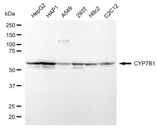
Western blotting analysis using CYP7B1 antibody. Total cell lysates (30 μg) from various cell lines were loaded and separated by SDS-PAGE. The blot was incubated with CYP7B1 antibody and HRP-conjugated goat anti-mouse secondary antibody respectively.ICC/IF
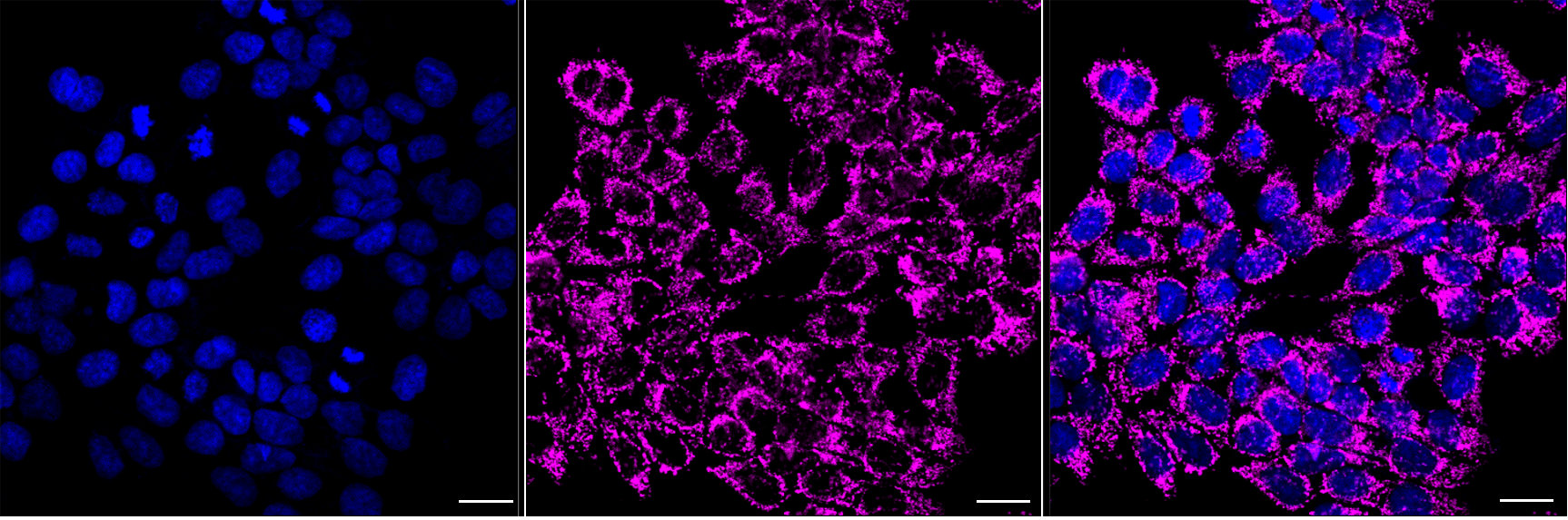
Immunocytochemical staining of HAP1 cells with CYP7B1 antibody. Nuclei were stained blue with DAPI; CYP7B1 stained magenta with Alexa 647. Images were taken using Leica stellaris 5. Protein abundance based on laser Intensity and smart gain: High. Scale bar, 20 μm.FC
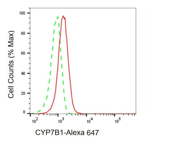
Flow cytometric analysis of CYP7B1 expression in HAP-1 cells using CYP7B1 antibody. Green, isotype control; red, CYP7B1.| Product Name | Mouse anti-CYP7B1 Monoclonal antibody |
|---|---|
| Antibody Type | Primary Antibodies |
| Immunogen | Recombinant Human CYP7B1 protein. |
| Clonality | Monoclonal |
|---|---|
| Isotype | IgG1 |
| Host Species | Mouse |
| Tested Applications | FCICC/IFWB |
| WB:1:200-1:1000 ICC/IF:1:100-1:1000 FC:1:200-1:2000 |
|
| Species Reactivity | HumanMouseRat |
| Concentration | 1mg/ml |
| Purification | Affinity purified |
| Gene Symbol | CYP7B1 |
|---|---|
| Gene Synonyms | CP7B CBAS3 SPG5A |
| Gene Full Name | cytochrome P450 family 7 subfamily B member 1 |
| Gene Summary | This gene encodes a member of the cytochrome P450 superfamily of enzymes. The cytochrome P450 proteins are monooxygenases which catalyze many reactions involved in drug metabolism and synthesis of cholesterol, steroids and other lipids. This endoplasmic reticulum membrane protein catalyzes the first reaction in the cholesterol catabolic pathway of extrahepatic tissues, which converts cholesterol to bile acids. This enzyme likely plays a minor role in total bile acid synthesis, but may also be involved in the development of atherosclerosis, neurosteroid metabolism and sex hormone synthesis. Mutations in this gene have been associated with hereditary spastic paraplegia (SPG5 or HSP), an autosomal recessive disorder. [provided by RefSeq, Apr 2016] |
| Alternative Names | CYP7B1 Cytochrome P450 Family 7 Subfamily B Member 1 Cytochrome P450 Subfamily VIIB (Oxysterol 7 Alpha-Hydroxylase) Polypeptide 1 25/26-Hydroxycholesterol 7-Alpha-Hydroxylase 24-Hydroxycholesterol 7-Alpha-Hydroxylase 3-Hydroxysteroid 7-Alpha Hydroxylase Oxysterol 7-Alpha-Hydroxylase Cytochrome P450 7B1 SPG5A Cytochrome P450 Family 7 Subfamily B Polypeptide 1 Spastic Paraplegia 5A (Autosomal Recessive) 25-Hydroxycholesterol 7-Alpha-Hydroxylase EC 1.14.14.26 EC 1.14.14.29 CBAS3 CP7B |
| Cellular Localization | Endoplasmic reticulum membrane; Multi-pass membrane protein. Microsome membrane; Multi-pass membrane protein. |
WB
Western blotting analysis using CYP7B1 antibody. Total cell lysates (30 μg) from various cell lines were loaded and separated by SDS-PAGE. The blot was incubated with CYP7B1 antibody and HRP-conjugated goat anti-mouse secondary antibody respectively.
ICC/IF
Immunocytochemical staining of HAP1 cells with CYP7B1 antibody. Nuclei were stained blue with DAPI; CYP7B1 stained magenta with Alexa 647. Images were taken using Leica stellaris 5. Protein abundance based on laser Intensity and smart gain: High. Scale bar, 20 μm.
FC
Flow cytometric analysis of CYP7B1 expression in HAP-1 cells using CYP7B1 antibody. Green, isotype control; red, CYP7B1.| Application Notes | WB:1:200-1:1000 ICC/IF:1:100-1:1000 FC:1:200-1:2000 |
|---|
| Form | Liquid |
|---|---|
| Storage Instructions | Upon receipt, store at -20°C or -80°C. Avoid repeated freeze. |
| Storage Buffer | PBS (pH 7.4) containing 50% glycerol, and 0.02% sodium azide. |


