WB
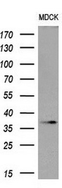
Western blot analysis of extracts (10ug) from 1 cell line by using anti-OCT4 monoclonal antibody at 1:200 dilution.WB
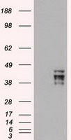
HEK293T cells were transfected with the pCMV6-ENTRY control (Left lane) or pCMV6-ENTRY OCT4 (Right lane) cDNA for 48 hrs and lysed. Equivalent amounts of cell lysates (5 ug per lane) were separated by SDS-PAGE and immunoblotted with anti-OCT4.IHC
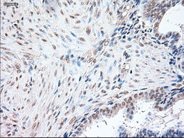
Immunohistochemical staining of paraffin-embedded Human endometrium tissue within the normal limits using anti-OCT4 mouse monoclonal antibody. (Heat-induced epitope retrieval by 10mM citric buffer, pH6.0, 100°C for 10min)ICC/IF
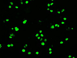
Anti-OCT4 mouse monoclonal antibody immunofluorescent staining of COS7 cells transiently transfected by pCMV6-ENTRY OCT4 .| Product Name | Mouse monoclonal antibody to OCT4/POU5F1 |
|---|---|
| Antibody Type | Primary Antibodies |
| Immunogen | Full length human recombinant protein of human POU5F1 (NP_002692) produced in E.coli. |
| Clonality | Monoclonal |
|---|---|
| Isotype | IgG1 |
| Host Species | Mouse |
| Tested Applications | ICC/IFIHCWB |
| WB:1:5000 IHC:1:50 ICC/IF:1:100 |
|
| Species Reactivity | Human |
| Concentration | 0.5mg/ml |
| Purification | Protein A/G |
| Gene Symbol | POU5F1 |
|---|---|
| Gene Synonyms | OCT3 OCT4 OTF3 OTF4 OTF-3 Oct-3 Oct-4 Oct3/4 OCT4Borf1 |
| Gene Full Name | POU class 5 homeobox 1 |
| Gene Summary | This gene encodes a transcription factor containing a POU homeodomain that plays a key role in embryonic development and stem cell pluripotency. Aberrant expression of this gene in adult tissues is associated with tumorigenesis. This gene can participate in a translocation with the Ewing's sarcoma gene on chromosome 21, which also leads to tumor formation. Alternative splicing, as well as usage of alternative AUG and non-AUG translation initiation codons, results in multiple isoforms. One of the AUG start codons is polymorphic in human populations. Related pseudogenes have been identified on chromosomes 1, 3, 8, 10, and 12. [provided by RefSeq, Oct 2013] |
| Alternative Names | Oct 3 Oct 4 OCT3 OCT3/4 OCT4 Octamer binding protein 3 Octamer binding protein 4 OTF 3 OTF3 OTF4 POU class 5 homeobox 1 POU5F1 |
| Molecular Weight(MW) | 39 kDa(Observed band size: 38.4 kDa) |
| Cellular Localization | Cytoplasm,Nucleus. |
WB
Western blot analysis of extracts (10ug) from 1 cell line by using anti-OCT4 monoclonal antibody at 1:200 dilution.
WB
HEK293T cells were transfected with the pCMV6-ENTRY control (Left lane) or pCMV6-ENTRY OCT4 (Right lane) cDNA for 48 hrs and lysed. Equivalent amounts of cell lysates (5 ug per lane) were separated by SDS-PAGE and immunoblotted with anti-OCT4.
IHC
Immunohistochemical staining of paraffin-embedded Human endometrium tissue within the normal limits using anti-OCT4 mouse monoclonal antibody. (Heat-induced epitope retrieval by 10mM citric buffer, pH6.0, 100°C for 10min)
ICC/IF
Anti-OCT4 mouse monoclonal antibody immunofluorescent staining of COS7 cells transiently transfected by pCMV6-ENTRY OCT4 .| Application Notes | WB:1:5000 IHC:1:50 ICC/IF:1:100 |
|---|
| Form | Liquid |
|---|---|
| Storage Instructions | Stable for 12 months from date of receipt. Store at -20°C as received. |
| Storage Buffer | PBS (pH 7.3) containing 1% BSA, 50% glycerol and 0.02% sodium azide. |


