WB
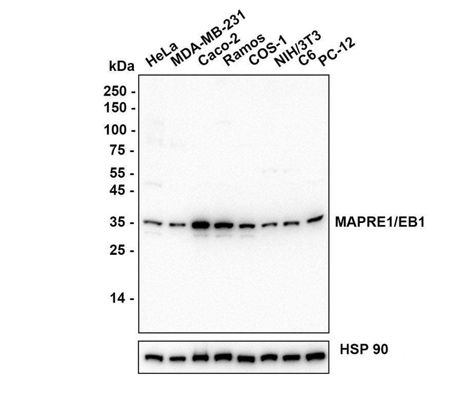
Western blot analysis of MAPRE1 / EB1 on different lysates with Rabbit anti-MAPRE1 / EB1 antibody at 1/5,000 dilution. Lysates/proteins at 10 µg/Lane. Exposure time: 25 seconds; Blocking: 5% NFDM/TBST, 1 hour at room temperature. Primary antibody: 1/5,000 in primary antibody dilution buffer, overnight at 4 °C. Secondary antibody: Goat anti-Rabbit IgG-HRP, 1/50,000 in 5% NFDM/TBST, 1 hour at room temperature.IHC
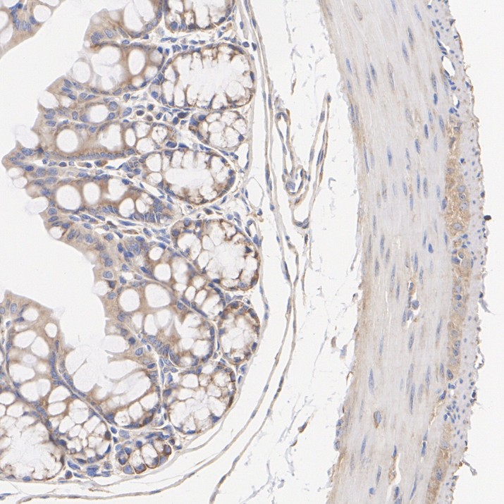
Application: Immunohistochemistry (IHC-P) Species: Mouse Tissue: Colon Sample: Paraffin-embedded section Antigen retrieval: Heat-mediated, Tris-EDTA buffer (pH 9.0), 20 minutes at 95°C. Wash buffer: 1× TBST Endogenous peroxidase blocking: 3% H₂O₂, 10 minutes at room temperature. Blocking: 1% BSA + 10% normal goat serum, 10 minutes at room temperature. Primary antibody: 1/1,000, 1 hour at room temperature. Secondary antibody: 20 minutes at room temperature.ICC/IF
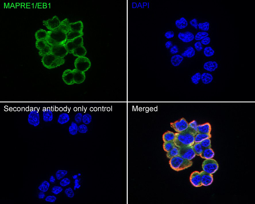
Application: Immunocytochemistry (IF-cell) Species: Rat Sample: PC-12 (Rat pheochromocytoma cell (undifferentiated)) Fixation: 4% Paraformaldehyde, 15 minutes at room temperature. Permeabilization: 0.1% Triton X-100, 15 minutes at room temperature. Blocking: 1% BSA + 10% normal goat serum, 1 hour at room temperature. Antibody dilution buffer: 1% BSA in PBST. Primary antibody: 1/500, overnight at 4°C. Secondary antibody: Goat Anti-Rabbit IgG (488), 45 minutes at room temperature. Counterstain: Beta tubulin (Red), 1/100, overnight at 4℃. The nuclear counterstain was DAPI (Blue).IP
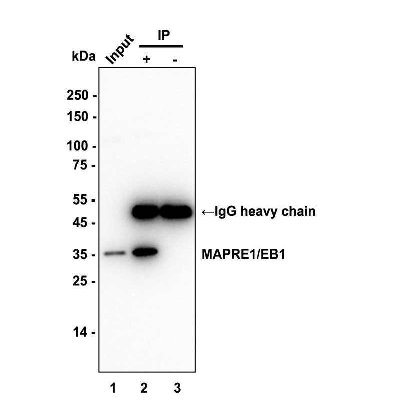
Immunoprecipitation (IP) MAPRE1 / EB1 was immunoprecipitated in 0.2 mg HeLa (Human cervix adenocarcinoma epithelial cell) cell lysate with Rabbit anti-MAPRE1 / EB1 antibody at 2 µg/10 µl beads. Western blot was performed from the immunoprecipitate using MAPRE1 / EB1 Recombinant Rabbit Monoclonal Antibody at 1/5,000 dilution. Alpaca anti-Rabbit IgG Fc, Recombinant VHH monoclonal Antibody at 1/50000 dilution was used for 1 hour at room temperature. Lane 1: HeLa cell lysate (input) Lane 2: Rabbit anti-MAPRE1 / EB1 antibody IP in HeLa cell lysate Lane 3: Rabbit IgG instead of Rabbit anti-MAPRE1 / EB1 antibody in HeLa cell lysate Exposure time: 10 seconds Blocking: 5% NFDM/TBST, 1 hour at room temperature Primary dilution: 1/5,000 in primary antibody dilution buffer, 2 hours at room temperatureFC
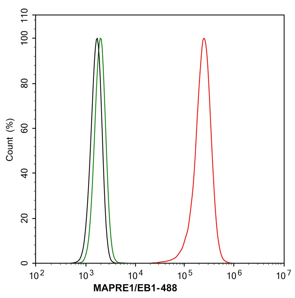
Application: Flow Cytometry (Intra) Species: Rat Sample: PC-12 (Rat pheochromocytoma cells (undifferentiated)) Fixation: 4% Paraformaldehyde, 15 minutes at room temperature. Permeabilization: 0.1% Tween-20, 15 minutes at room temperature. Blocking: 1% BSA + 10% normal goat serum, 15 minutes at room temperature. Antibody dilution buffer: 1x PBS. Primary antibody: MAPRE1 / EB1 Recombinant Rabbit Monoclonal Antibody (1/1,000, Red) compared with Rabbit IgG Isotype Control (Green), 15 minutes at room temperature. Secondary antibody: Goat Anti-Rabbit IgG (488), 15 minutes at room temperature.| Product Name | MAPRE1 / EB1 Recombinant Rabbit Monoclonal Antibody |
|---|---|
| Antibody Type | Primary Antibodies |
| Immunogen | Recombinant protein within human MAPRE1 aa 101-250. |
| Clonality | Monoclonal |
|---|---|
| Isotype | IgG |
| Host Species | Rabbit |
| Tested Applications | FCICC/IFIHCIPWB |
| WB:1:5000 IHC:1:1000 ICC/IF:1:500 FC: 1:1000 IP:1-2μg/sample |
|
| Species Reactivity | HumanMonkeyMousePigRat |
| Concentration | 1mg/ml |
| Purification | Protein A |
| Gene Symbol | MT-ND1 |
|---|---|
| Gene Synonyms | MTND1 ND1 |
| Gene Full Name | mitochondrially encoded NADH dehydrogenase 1 |
| Gene Summary | Enables NADH dehydrogenase (ubiquinone) activity. Involved in mitochondrial electron transport, NADH to ubiquinone and mitochondrial respiratory chain complex I assembly. Located in mitochondrial inner membrane. Part of respiratory chain complex I. Implicated in several diseases, including MELAS syndrome; neurodegenerative disease (multiple); optic nerve disease (multiple); toxic shock syndrome; and type 2 diabetes mellitus. Biomarker of Alzheimer's disease; Parkinson's disease; and multiple sclerosis. [provided by Alliance of Genome Resources, Jul 2025] |
| Alternative Names | Microtubule-associated protein RP/EB family member 1 APC-binding protein EB1 End-binding protein 1 (EB1) MAPRE1 |
| Molecular Weight(MW) | 30 kDa(Observed band size:30-35 kDa ) |
| Cellular Localization | Cytoplasm, cytoskeleton, microtubule organizing center, centrosome, Golgi apparatus, spindle, spindle pole, Cell membrane. |
WB
Western blot analysis of MAPRE1 / EB1 on different lysates with Rabbit anti-MAPRE1 / EB1 antibody at 1/5,000 dilution. Lysates/proteins at 10 µg/Lane. Exposure time: 25 seconds; Blocking: 5% NFDM/TBST, 1 hour at room temperature. Primary antibody: 1/5,000 in primary antibody dilution buffer, overnight at 4 °C. Secondary antibody: Goat anti-Rabbit IgG-HRP, 1/50,000 in 5% NFDM/TBST, 1 hour at room temperature.
IHC
Application: Immunohistochemistry (IHC-P) Species: Mouse Tissue: Colon Sample: Paraffin-embedded section Antigen retrieval: Heat-mediated, Tris-EDTA buffer (pH 9.0), 20 minutes at 95°C. Wash buffer: 1× TBST Endogenous peroxidase blocking: 3% H₂O₂, 10 minutes at room temperature. Blocking: 1% BSA + 10% normal goat serum, 10 minutes at room temperature. Primary antibody: 1/1,000, 1 hour at room temperature. Secondary antibody: 20 minutes at room temperature.
ICC/IF
Application: Immunocytochemistry (IF-cell) Species: Rat Sample: PC-12 (Rat pheochromocytoma cell (undifferentiated)) Fixation: 4% Paraformaldehyde, 15 minutes at room temperature. Permeabilization: 0.1% Triton X-100, 15 minutes at room temperature. Blocking: 1% BSA + 10% normal goat serum, 1 hour at room temperature. Antibody dilution buffer: 1% BSA in PBST. Primary antibody: 1/500, overnight at 4°C. Secondary antibody: Goat Anti-Rabbit IgG (488), 45 minutes at room temperature. Counterstain: Beta tubulin (Red), 1/100, overnight at 4℃. The nuclear counterstain was DAPI (Blue).
IP
Immunoprecipitation (IP) MAPRE1 / EB1 was immunoprecipitated in 0.2 mg HeLa (Human cervix adenocarcinoma epithelial cell) cell lysate with Rabbit anti-MAPRE1 / EB1 antibody at 2 µg/10 µl beads. Western blot was performed from the immunoprecipitate using MAPRE1 / EB1 Recombinant Rabbit Monoclonal Antibody at 1/5,000 dilution. Alpaca anti-Rabbit IgG Fc, Recombinant VHH monoclonal Antibody at 1/50000 dilution was used for 1 hour at room temperature. Lane 1: HeLa cell lysate (input) Lane 2: Rabbit anti-MAPRE1 / EB1 antibody IP in HeLa cell lysate Lane 3: Rabbit IgG instead of Rabbit anti-MAPRE1 / EB1 antibody in HeLa cell lysate Exposure time: 10 seconds Blocking: 5% NFDM/TBST, 1 hour at room temperature Primary dilution: 1/5,000 in primary antibody dilution buffer, 2 hours at room temperature
FC
Application: Flow Cytometry (Intra) Species: Rat Sample: PC-12 (Rat pheochromocytoma cells (undifferentiated)) Fixation: 4% Paraformaldehyde, 15 minutes at room temperature. Permeabilization: 0.1% Tween-20, 15 minutes at room temperature. Blocking: 1% BSA + 10% normal goat serum, 15 minutes at room temperature. Antibody dilution buffer: 1x PBS. Primary antibody: MAPRE1 / EB1 Recombinant Rabbit Monoclonal Antibody (1/1,000, Red) compared with Rabbit IgG Isotype Control (Green), 15 minutes at room temperature. Secondary antibody: Goat Anti-Rabbit IgG (488), 15 minutes at room temperature.| Application Notes | WB:1:5000 IHC:1:1000 ICC/IF:1:500 FC: 1:1000 IP:1-2μg/sample |
|---|
| Form | Liquid |
|---|---|
| Storage Instructions | Shipped at 4℃. Store at +4℃ short term (1-2 weeks). Store at -20℃ long term. |
| Storage Buffer | 1*PBS (pH7.4), 0.1% BSA, 40% Glycerol, 0.2% Proclean 950. |


