WB
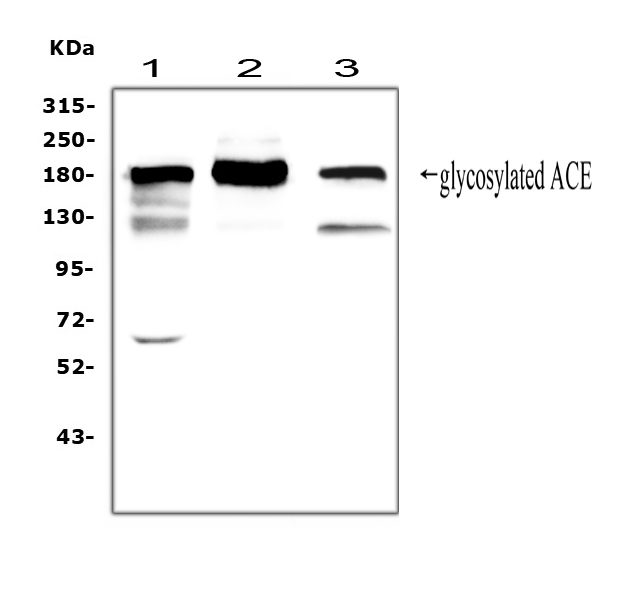
Western blot analysis of ACE using anti-ACE antibody. The sample well of each lane was loaded with 30 ug of sample under reducing conditions. Lane 1: rat lung tissue lysates, Lane 2: mouse lung tissue lysates, Lane 3: human Raji whole cell lysates. After electrophoresis, proteins were transferred to a membrane. Then the membrane was incubated with rabbit anti-ACE antigen affinity purified polyclonal antibody at a dilution of 1:1000 and probed with a goat anti-rabbit IgG-HRP secondary antibody. The signal is developed using ECL Plus Western Blotting Substrate.IHC
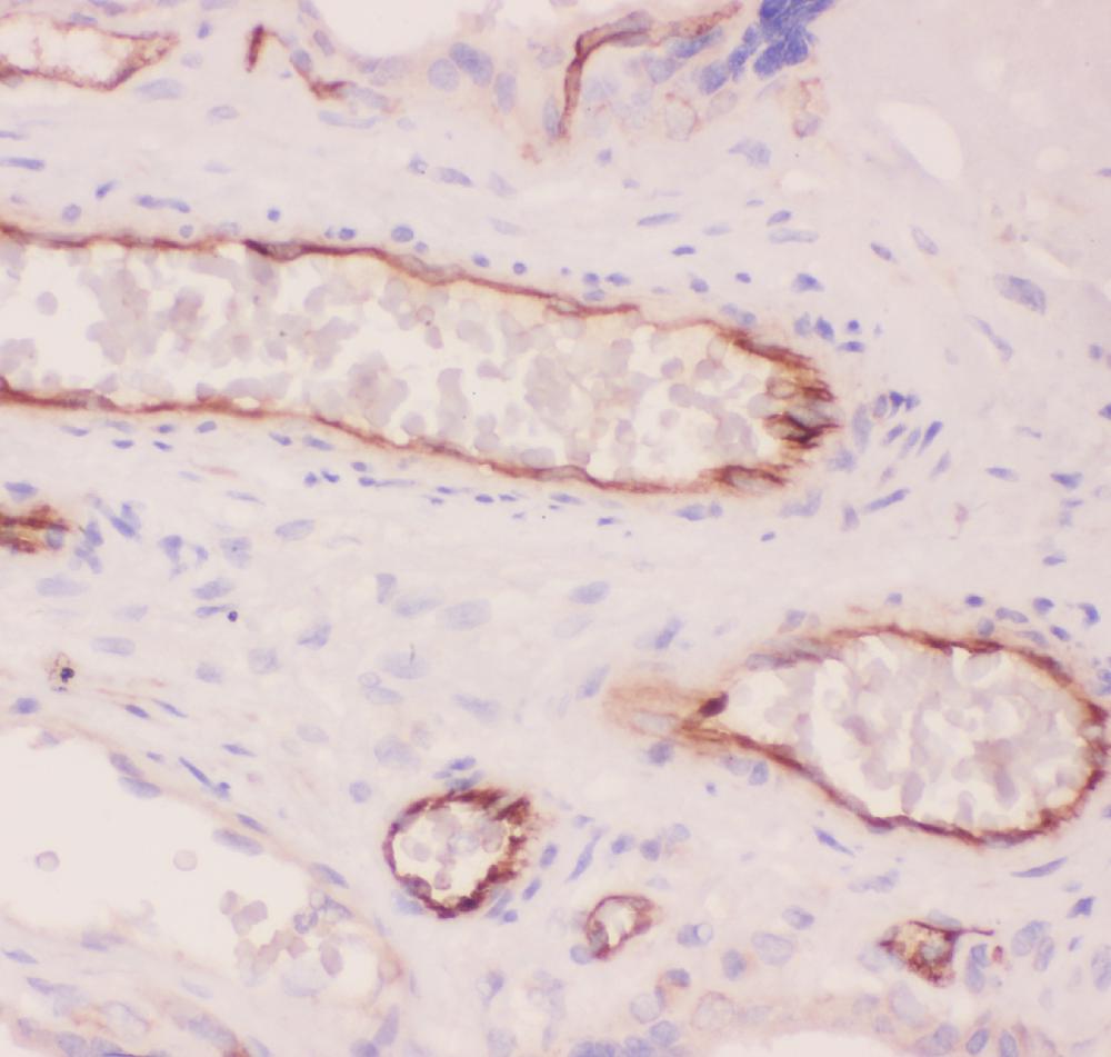
IHC analysis of ACE using anti-ACE antibody. ACE was detected in a paraffin-embedded section of human placenta tissue. Biotinylated goat anti-rabbit IgG was used as secondary antibody. The tissue section was incubated with rabbit anti-ACE Antibody at a dilution of 1:200 and developed using Strepavidin-Biotin-Complex (SABC) with DAB as the chromogen.IHC
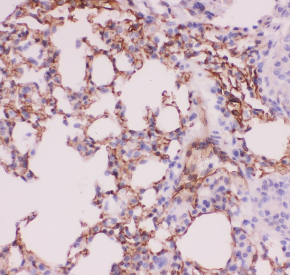
IHC analysis of ACE using anti-ACE antibody. ACE was detected in a paraffin-embedded section of mouse lung tissue. Biotinylated goat anti-rabbit IgG was used as secondary antibody. The tissue section was incubated with rabbit anti-ACE Antibody at a dilution of 1:200 and developed using Strepavidin-Biotin-Complex (SABC) with DAB as the chromogen.IHC
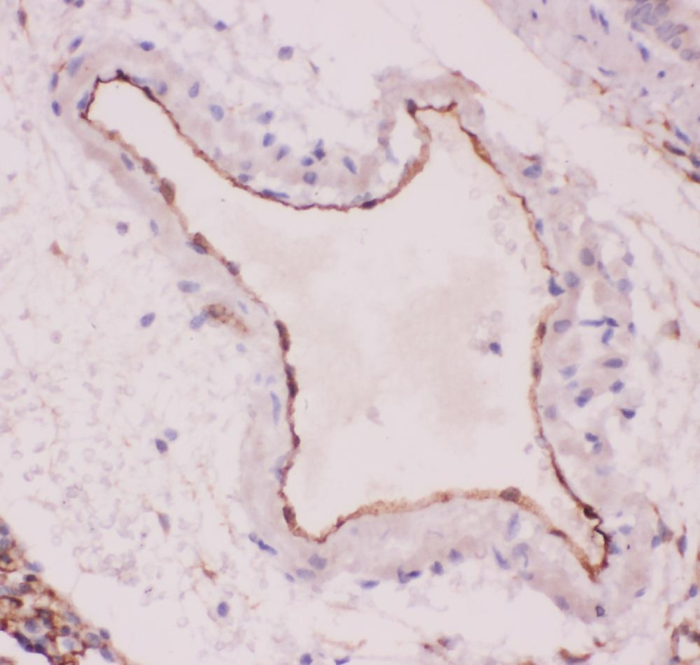
IHC analysis of ACE using anti-ACE antibody. ACE was detected in a paraffin-embedded section of rat lung tissue. Biotinylated goat anti-rabbit IgG was used as secondary antibody. The tissue section was incubated with rabbit anti-ACE Antibody at a dilution of 1:200 and developed using Strepavidin-Biotin-Complex (SABC) with DAB as the chromogen.ICC/IF
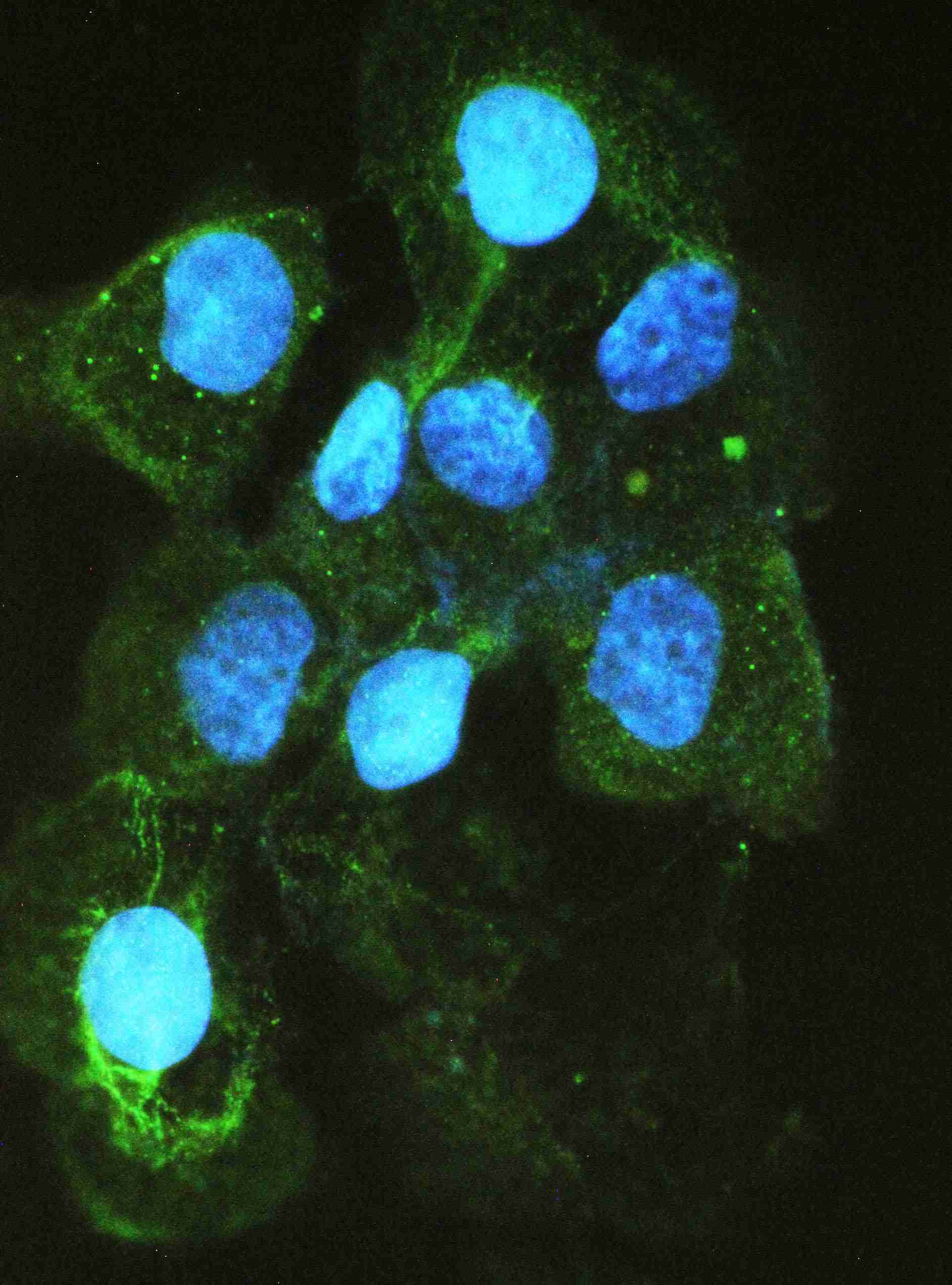
ICC/IF analysis of ACE using anti- ACE antibody. ACE was detected in immunocytochemical section of A431 cell. Enzyme antigen retrieval was performed using IHC enzyme antigen retrieval reagent for 15 mins. The cells were blocked with 10% goat serum. And then incubated with 2μg/mL rabbit anti- ACE Antibody overnight at 4°C. Fluoro488 Conjugated Goat Anti-Rabbit IgG was used as secondary antibody at 1:100 dilution and incubated for 30 minutes at 37°C. The section was counterstained with DAPI. Visualize using a fluorescence microscope and filter sets appropriate for the label used.| Product Name | Rabbit polyclonal antibody to ACE |
|---|---|
| Antibody Type | Primary Antibodies |
| Immunogen | E.coli-derived human ACE recombinant protein (Position: K651-Y864). Human ACE shares 73% and 76% amino acid (aa) sequences identity with mouse and rat ACE, respectively. |
| Clonality | Polyclonal |
|---|---|
| Isotype | IgG |
| Host Species | Rabbit |
| Tested Applications | ICC/IFIHCWB |
| WB:1:500-1:2000 IHC:1:50-1:400 ICC/IF:1:50-1:400 |
|
| Species Reactivity | HumanMouseRat |
| Concentration | 0.5mg/ml |
| Purification | Affinity purified |
| Gene Symbol | ACE |
|---|---|
| Gene Synonyms | DCP ACE1 DCP1 CD143 |
| Gene Full Name | angiotensin I converting enzyme |
| Gene Summary | This gene encodes an enzyme involved in blood pressure regulation and electrolyte balance. It catalyzes the conversion of angiotensin I into a physiologically active peptide angiotensin II. Angiotensin II is a potent vasopressor and aldosterone-stimulating peptide that controls blood pressure and fluid-electrolyte balance. This angiotensin converting enzyme (ACE) also inactivates the vasodilator protein, bradykinin. Accordingly, the encoded enzyme increases blood pressure and is a drug target of ACE inhibitors, which are often prescribed to reduce blood pressure. This enzyme additionally plays a role in fertility through its ability to cleave and release GPI-anchored membrane proteins in spermatozoa. Many studies have associated the presence or absence of a 287 bp Alu repeat element in this gene with the levels of circulating enzyme. This polymorphism, as well as mutations in this gene, have been implicated in a wide variety of diseases including cardiovascular pathophysiologies, psoriasis, renal disease, stroke, and Alzheimer's disease. Regulation of the homologous ACE2 gene may be involved in progression of disease caused by several human coronaviruses, including SARS-CoV and SARS-CoV-2. Alternative splicing results in multiple transcript variants encoding both somatic (sACE) and male-specific testicular (tACE) isoforms. [provided by RefSeq, Sep 2020] |
| Alternative Names | ACE ACE1 Angiotensin converting enzyme CD143 DCP DCP1 Dipeptidyl carboxypeptidase I Kininase II MVCD3 |
| Molecular Weight(MW) | 54 kDa(Observed band size:150-180 kDa) |
| Cellular Localization | Cell membrane,Cytoplasm,Membrane,Secreted. |
WB
Western blot analysis of ACE using anti-ACE antibody. The sample well of each lane was loaded with 30 ug of sample under reducing conditions. Lane 1: rat lung tissue lysates, Lane 2: mouse lung tissue lysates, Lane 3: human Raji whole cell lysates. After electrophoresis, proteins were transferred to a membrane. Then the membrane was incubated with rabbit anti-ACE antigen affinity purified polyclonal antibody at a dilution of 1:1000 and probed with a goat anti-rabbit IgG-HRP secondary antibody. The signal is developed using ECL Plus Western Blotting Substrate.
IHC
IHC analysis of ACE using anti-ACE antibody. ACE was detected in a paraffin-embedded section of human placenta tissue. Biotinylated goat anti-rabbit IgG was used as secondary antibody. The tissue section was incubated with rabbit anti-ACE Antibody at a dilution of 1:200 and developed using Strepavidin-Biotin-Complex (SABC) with DAB as the chromogen.
IHC
IHC analysis of ACE using anti-ACE antibody. ACE was detected in a paraffin-embedded section of mouse lung tissue. Biotinylated goat anti-rabbit IgG was used as secondary antibody. The tissue section was incubated with rabbit anti-ACE Antibody at a dilution of 1:200 and developed using Strepavidin-Biotin-Complex (SABC) with DAB as the chromogen.
IHC
IHC analysis of ACE using anti-ACE antibody. ACE was detected in a paraffin-embedded section of rat lung tissue. Biotinylated goat anti-rabbit IgG was used as secondary antibody. The tissue section was incubated with rabbit anti-ACE Antibody at a dilution of 1:200 and developed using Strepavidin-Biotin-Complex (SABC) with DAB as the chromogen.
ICC/IF
ICC/IF analysis of ACE using anti- ACE antibody. ACE was detected in immunocytochemical section of A431 cell. Enzyme antigen retrieval was performed using IHC enzyme antigen retrieval reagent for 15 mins. The cells were blocked with 10% goat serum. And then incubated with 2μg/mL rabbit anti- ACE Antibody overnight at 4°C. Fluoro488 Conjugated Goat Anti-Rabbit IgG was used as secondary antibody at 1:100 dilution and incubated for 30 minutes at 37°C. The section was counterstained with DAPI. Visualize using a fluorescence microscope and filter sets appropriate for the label used.| Application Notes | WB:1:500-1:2000 IHC:1:50-1:400 ICC/IF:1:50-1:400 |
|---|
| Form | Liquid |
|---|---|
| Storage Instructions | 12 months from date of receipt,-20℃ as supplied. |
| Storage Buffer | 500 ug/ml antibody with PBS, 0.02% NaN3, 1 mg/ml BSA and 50% glycerol. |


