WB
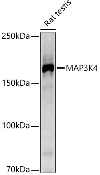
Western blot analysis of lysates from Rat testis, using MAP3K4 Rabbit mAb at 1:1000 dilution. Secondary antibody: HRP-conjugated Goat anti-Rabbit IgG (H+L) at 1:10000 dilution. Lysates/proteins: 25μg per lane. Blocking buffer: 3% nonfat dry milk in TBST. Detection: ECL Basic Kit . Exposure time: 180s.IHC
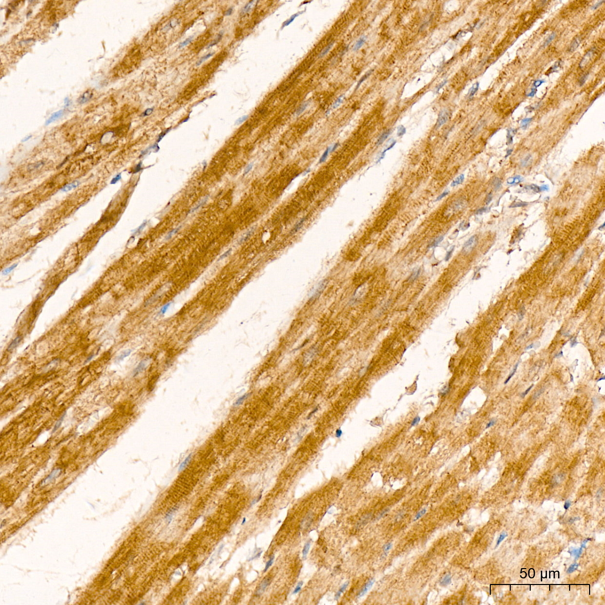
Immunohistochemistry analysis of paraffin embedded Rat heart tissue using MAP3K4 Rabbit mAb at a dilution of 1:200 (40x lens). High pressure antigen retrieval was performed with 0.01 M citrate buffer (pH 6.0) prior to IHC staining.IHC
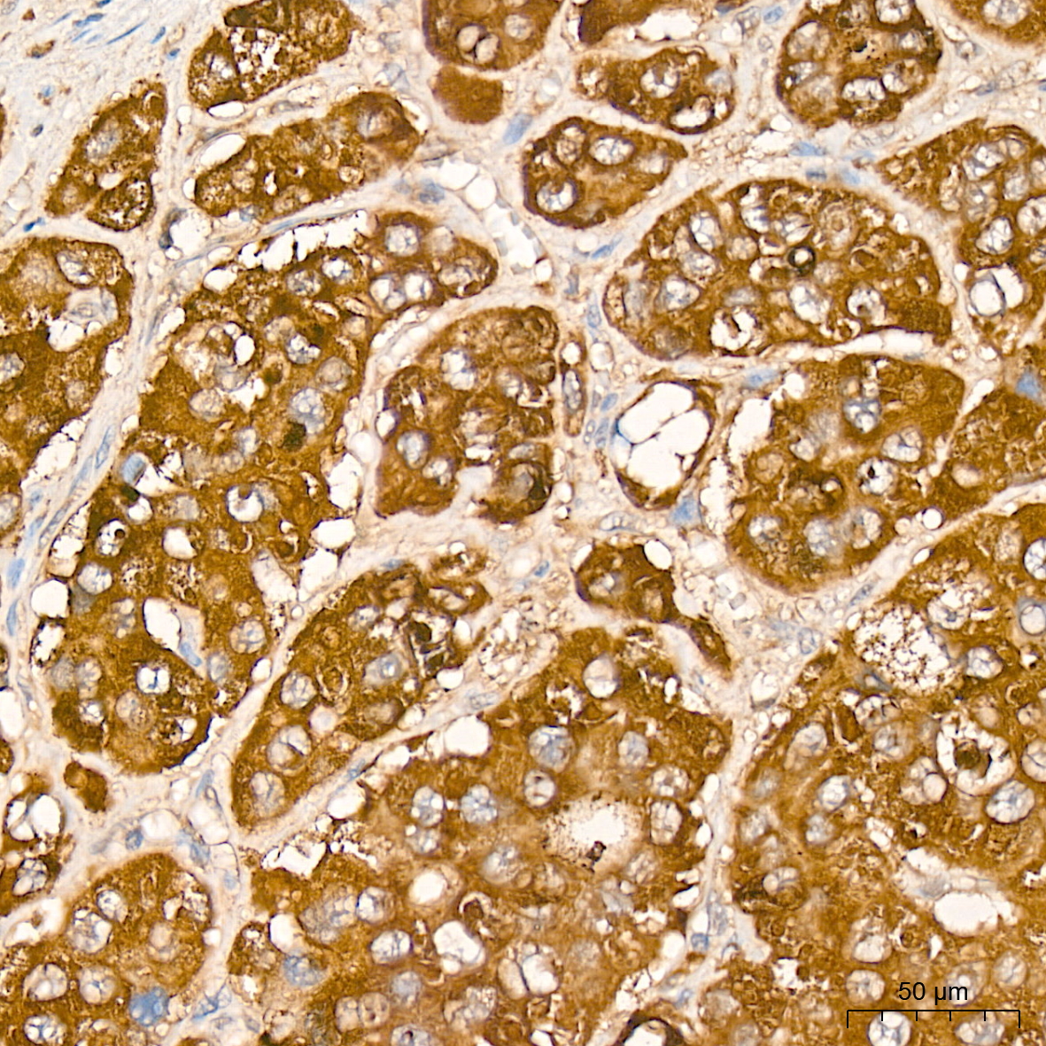
Immunohistochemistry analysis of paraffin embedded Human liver cancer tissue using MAP3K4 Rabbit mAb at a dilution of 1:200 (40x lens). High pressure antigen retrieval was performed with 0.01 M citrate buffer (pH 6.0) prior to IHC staining.IHC
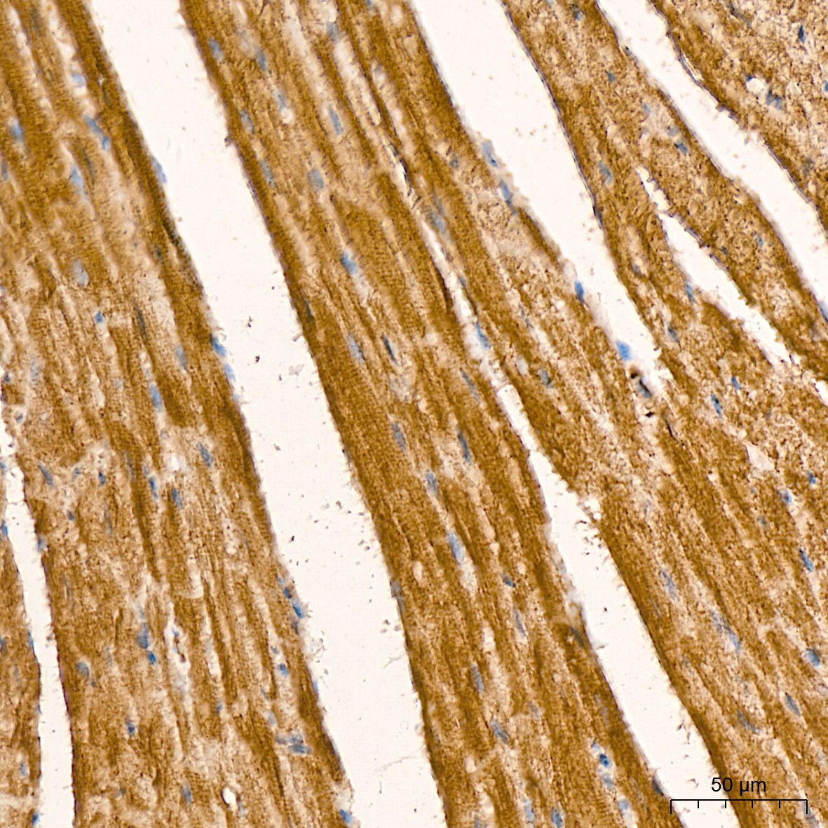
Immunohistochemistry analysis of paraffin embedded Mouse heart tissue using MAP3K4 Rabbit mAb at a dilution of 1:200 (40x lens). High pressure antigen retrieval was performed with 0.01 M citrate buffer (pH 6.0) prior to IHC staining.| Product Name | MAP3K4 Rabbit mAb |
|---|---|
| Antibody Type | Primary Antibodies |
| Immunogen | Synthetic peptide. This information is considered to be commercially sensitive. |
| Clonality | Monoclonal |
|---|---|
| Isotype | IgG |
| Host Species | Rabbit |
| Tested Applications | IHCWB |
| WB:1:500-1:1000 IHC:1:50 - 1:200 |
|
| Species Reactivity | HumanMouseRat |
| Concentration | 1mg/ml |
| Purification | Affinity purified |
| Gene Symbol | MAP3K4 |
|---|---|
| Gene Synonyms | MTK1 MEKK4 MEKK 4 MAPKKK4 PRO0412 |
| Gene Full Name | mitogen-activated protein kinase kinase kinase 4 |
| Gene Summary | The central core of each mitogen-activated protein kinase (MAPK) pathway is a conserved cascade of 3 protein kinases: an activated MAPK kinase kinase (MAPKKK) phosphorylates and activates a specific MAPK kinase (MAPKK), which then activates a specific MAPK. While the ERK MAPKs are activated by mitogenic stimulation, the CSBP2 and JNK MAPKs are activated by environmental stresses such as osmotic shock, UV irradiation, wound stress, and inflammatory factors. This gene encodes a MAPKKK, the MEKK4 protein, also called MTK1. This protein contains a protein kinase catalytic domain at the C terminus. The N-terminal nonkinase domain may contain a regulatory domain. Expression of MEKK4 in mammalian cells activated the CSBP2 and JNK MAPK pathways, but not the ERK pathway. In vitro kinase studies indicated that recombinant MEKK4 can specifically phosphorylate and activate PRKMK6 and SERK1, MAPKKs that activate CSBP2 and JNK, respectively but cannot phosphorylate PRKMK1, an MAPKK that activates ERKs. MEKK4 is a major mediator of environmental stresses that activate the CSBP2 MAPK pathway, and a minor mediator of the JNK pathway. Several alternatively spliced transcripts encoding distinct isoforms have been described. [provided by RefSeq, May 2014] |
| Alternative Names | MTK1 MEKK4 MEKK 4 MAPKKK4 PRO0412 MAP3K4 |
| Molecular Weight(MW) | 182 kDa(Observed band size:180 kDa) |
| Cellular Localization | cytoplasm, perinuclear region of cytoplasm. |
WB
Western blot analysis of lysates from Rat testis, using MAP3K4 Rabbit mAb at 1:1000 dilution. Secondary antibody: HRP-conjugated Goat anti-Rabbit IgG (H+L) at 1:10000 dilution. Lysates/proteins: 25μg per lane. Blocking buffer: 3% nonfat dry milk in TBST. Detection: ECL Basic Kit . Exposure time: 180s.
IHC
Immunohistochemistry analysis of paraffin embedded Rat heart tissue using MAP3K4 Rabbit mAb at a dilution of 1:200 (40x lens). High pressure antigen retrieval was performed with 0.01 M citrate buffer (pH 6.0) prior to IHC staining.
IHC
Immunohistochemistry analysis of paraffin embedded Human liver cancer tissue using MAP3K4 Rabbit mAb at a dilution of 1:200 (40x lens). High pressure antigen retrieval was performed with 0.01 M citrate buffer (pH 6.0) prior to IHC staining.
IHC
Immunohistochemistry analysis of paraffin embedded Mouse heart tissue using MAP3K4 Rabbit mAb at a dilution of 1:200 (40x lens). High pressure antigen retrieval was performed with 0.01 M citrate buffer (pH 6.0) prior to IHC staining.| Application Notes | WB:1:500-1:1000 IHC:1:50 - 1:200 |
|---|
| Form | Liquid |
|---|---|
| Storage Instructions | Store at -20℃. Avoid freeze / thaw cycles. |
| Storage Buffer | PBS containing 50% glycerol and 0.05% BSA, preserved with proclin300 or sodium azide (as specified on the Certificate of Analysis), pH 7.3. |


