WB
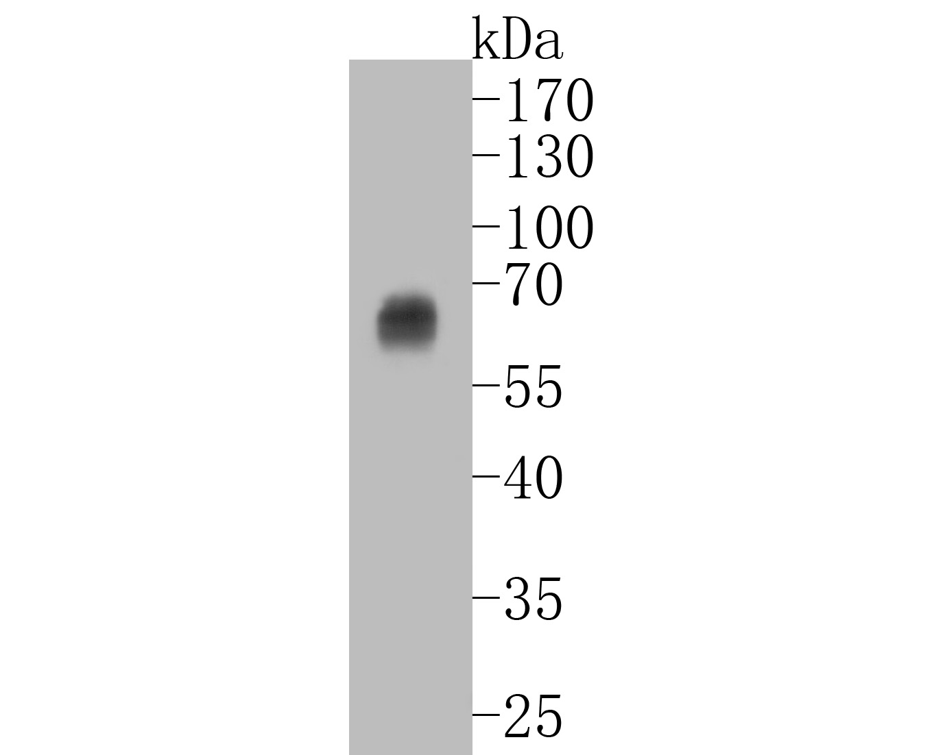
Western blot analysis of Oct-2 on Daudi cell lysates. Proteins were transferred to a PVDF membrane and blocked with 5% BSA in PBS for 1 hour at room temperature. The primary antibody (1/500) was used in 5% BSA at room temperature for 2 hours. Goat Anti-Rabbit IgG - HRP Secondary Antibody at 1:5,000 dilution was used for 1 hour at room temperature.IHC
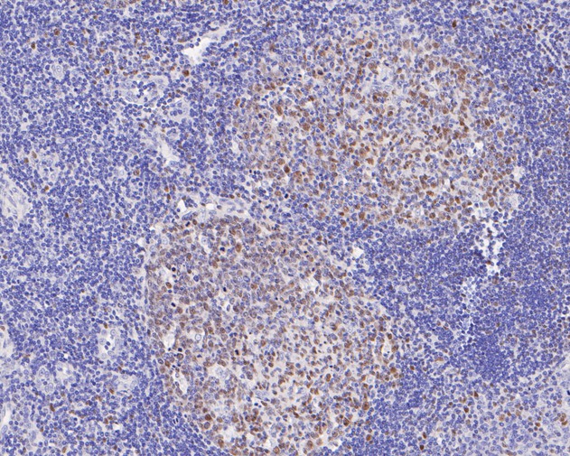
Immunohistochemical analysis of paraffin-embedded human tonsil tissue using anti-Oct-2 antibody. The section was pre-treated using heat mediated antigen retrieval with sodium citrate buffer (pH 6.0) (high pressure) for 2 minutes. The tissues were blocked in 5% BSA for 30 minutes at room temperature, washed with ddH2O and PBS, and then probed with the primary antibody (1/50) for 30 minutes at room temperature. The detection was performed using an HRP conjugated compact polymer system. DAB was used as the chromogen. Tissues were counterstained with hematoxylin and mounted with DPX.FC
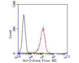
Flow cytometric analysis of Oct-2 was done on Daudi cells. The cells were fixed, permeabilized and stained with the primary antibody (1/50) (red). After incubation of the primary antibody at room temperature for an hour, the cells were stained with a Alexa Fluor 488-conjugated Goat anti-Rabbit IgG Secondary antibody at 1/1000 dilution for 30 minutes.Unlabelled sample was used as a control (cells without incubation with primary antibody; black).| Product Name | Oct-2 Recombinant Rabbit Monoclonal Antibody |
|---|---|
| Antibody Type | Primary Antibodies |
| Immunogen | Recombinant full length protein of human Oct-2. |
| Clonality | Monoclonal |
|---|---|
| Isotype | IgG |
| Host Species | Rabbit |
| Tested Applications | FCIHCWB |
| WB:1:500-1:1000 IHC:1:50-1:100 FC:1:50-1:100 |
|
| Species Reactivity | Human |
| Concentration | 1mg/ml |
| Purification | Protein A |
| Gene Symbol | POU2F2 |
|---|---|
| Gene Synonyms | OCT2 OTF2 Oct-2 |
| Gene Full Name | POU class 2 homeobox 2 |
| Gene Summary | The protein encoded by this gene is a homeobox-containing transcription factor of the POU domain family. The encoded protein binds the octamer sequence 5'-ATTTGCAT-3', a common transcription factor binding site in immunoglobulin gene promoters. Several transcript variants encoding different isoforms have been found for this gene. [provided by RefSeq, Oct 2011] |
| Alternative Names | class 2 antibody Lymphoid restricted immunoglobulin octamer binding protein antibody Lymphoid-restricted immunoglobulin octamer-binding protein NF-A2 antibody NF A2 antibody Oct 2 antibody Oct-2 antibody Octamer binding transcription factor 2 antibody Octamer-binding protein 2 antibody Octamer-binding transcription factor 2 antibody OTF-2 antibody OTF2 antibody PO2F2_HUMAN antibody POU domain antibody POU domain class 2 transcription factor 2 antibody POU2F2 antibody transcription factor 2 antibody |
| Molecular Weight(MW) | 51 kDa |
| Cellular Localization | Nucleus, Cytoplasm. |
WB
Western blot analysis of Oct-2 on Daudi cell lysates. Proteins were transferred to a PVDF membrane and blocked with 5% BSA in PBS for 1 hour at room temperature. The primary antibody (1/500) was used in 5% BSA at room temperature for 2 hours. Goat Anti-Rabbit IgG - HRP Secondary Antibody at 1:5,000 dilution was used for 1 hour at room temperature.
IHC
Immunohistochemical analysis of paraffin-embedded human tonsil tissue using anti-Oct-2 antibody. The section was pre-treated using heat mediated antigen retrieval with sodium citrate buffer (pH 6.0) (high pressure) for 2 minutes. The tissues were blocked in 5% BSA for 30 minutes at room temperature, washed with ddH2O and PBS, and then probed with the primary antibody (1/50) for 30 minutes at room temperature. The detection was performed using an HRP conjugated compact polymer system. DAB was used as the chromogen. Tissues were counterstained with hematoxylin and mounted with DPX.
FC
Flow cytometric analysis of Oct-2 was done on Daudi cells. The cells were fixed, permeabilized and stained with the primary antibody (1/50) (red). After incubation of the primary antibody at room temperature for an hour, the cells were stained with a Alexa Fluor 488-conjugated Goat anti-Rabbit IgG Secondary antibody at 1/1000 dilution for 30 minutes.Unlabelled sample was used as a control (cells without incubation with primary antibody; black).| Application Notes | WB:1:500-1:1000 IHC:1:50-1:100 FC:1:50-1:100 |
|---|
| Form | Liquid |
|---|---|
| Storage Instructions | Shipped at 4℃. Store at +4℃ short term (1-2 weeks). It is recommended to aliquot into single-use upon delivery. Store at -20℃ long term. |
| Storage Buffer | TBS (pH7.4), 0.05% BSA, 40% Glycerol. Preservative: 0.05% Sodium Azide. |


