WB
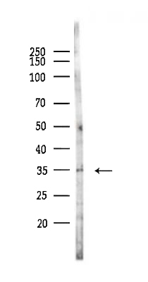
Western blot analysis of lysates from 293 cells(UV,30min treatment), using Phospho-TMEM173/STING (Ser366) Antibody at 1/1000 dilution. 5ug/NC membrane strip. Exposure for 2s with ECL Kit. Bands result from membrane strip incubation.IHC
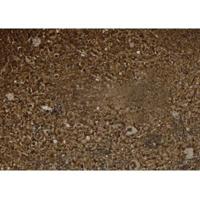
Rabbit polyclonal antibody to Phospho-TMEM173/STING at 1/100 staining mouse spleen tissue sections by IHC-P. The tissue was formaldehyde fixed and a heat mediated antigen retrieval step in citrate buffer was performed. The tissue was then blocked and incubated with the antibody for 1.5 hours at 22°C. An HRP conjugated goat anti-rabbit antibody was used as the secondary antibody.IHC
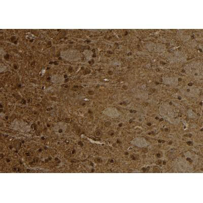
Rabbit polyclonal antibody to Phospho-TMEM173/STING at 1/100 staining rat brain tissue sections by IHC-P. The tissue was formaldehyde fixed and a heat mediated antigen retrieval step in citrate buffer was performed. The tissue was then blocked and incubated with the antibody for 1.5 hours at 22°C. An HRP conjugated goat anti-rabbit antibody was used as the secondary antibody.IHC
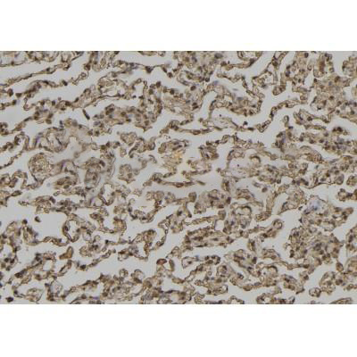
Rabbit polyclonal antibody to Phospho-TMEM173/STING at 1/100 staining human lung cancer tissue sections by IHC-P. The tissue was formaldehyde fixed and a heat mediated antigen retrieval step in citrate buffer was performed. The tissue was then blocked and incubated with the antibody for 1.5 hours at 22°C. An HRP conjugated goat anti-rabbit antibody was used as the secondary antibody.ICC/IF
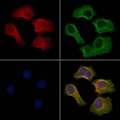
Rabbit polyclonal antibody to Phospho-TMEM173/STING staining HepG2 cells(4h of LPS treatment) by IF/ICC. The samples were fixed with PFA and permeabilized in 0.1% Triton X-100,then blocked in 10% serum for 45 minutes at 25°C. Samples were then incubated with primary Ab(1:200) and mouse anti-beta tubulin Ab for 1 hour at 37°C. An AlexaFluor594 conjugated goat anti-rabbit IgG(H+L) Ab(Red) and an AlexaFluor488 conjugated goat anti-mouse IgG(H+L) Ab(Green) were used as the secondary antibody. The nuclear counter stain is DAPI(blue).| Product Name | Rabbit polyclonal antibody to Phospho-TMEM173/STING (Ser366) |
|---|---|
| Antibody Type | Primary Antibodies |
| Immunogen | A synthesized peptide derived from human TMEM173/STING around the phosphorylation site of Ser366. |
| Modification | p-Ser366 |
| Clonality | Polyclonal |
|---|---|
| Isotype | IgG |
| Host Species | Rabbit |
| Tested Applications | ICC/IFIHCWB |
| WB:1:500-1:2000 IHC:1:50-1:200 ICC/IF:1:100-1:500 |
|
| Species Reactivity | HumanMouseRat |
| Concentration | 1mg/ml |
| Purification | Affinity purified |
| Gene Symbol | STING1 |
|---|---|
| Gene Synonyms | ERIS MITA MPYS SAVI NET23 STING hMITA hSTING TMEM173 STING-beta |
| Gene Full Name | stimulator of interferon response cGAMP interactor 1 |
| Gene Summary | This gene encodes a five transmembrane protein that functions as a major regulator of the innate immune response to viral and bacterial infections. The encoded protein is a pattern recognition receptor that detects cytosolic nucleic acids and transmits signals that activate type I interferon responses. The encoded protein has also been shown to play a role in apoptotic signaling by associating with type II major histocompatibility complex. Mutations in this gene are the cause of infantile-onset STING-associated vasculopathy. Alternate splicing results in multiple transcript variants. [provided by RefSeq, Sep 2014] |
| Alternative Names | endoplasmic reticulum IFN stimulator Endoplasmic reticulum interferon stimulator ERIS FLJ38577 hMITA hSTING Mediator of IRF3 activation MITA Mitochondrial mediator of IRF3 activation MPYS N terminal methionine proline tyrosine serine plasma membrane tetraspanner NET23 Stimulator of interferon genes Stimulator of interferon genes protein STING TM173_HUMAN Tmem173 Transmembrane protein 173 |
| Molecular Weight(MW) | 42 kDa(Observed band size:35-40kDa) |
| Cellular Localization | Endoplasmic reticulum membrane>Multi-pass membrane protein. Cytoplasm>Perinuclear region. Endoplasmic reticulum-Golgi intermediate compartment membrane>Multi-pass membrane protein. Cytoplasmic vesicle>Autophagosome membrane>Multi-pass membrane protein. Mitochondrion outer membrane>Multi-pass membrane protein. Cell membrane>Multi-pass membrane protein. |
WB
Western blot analysis of lysates from 293 cells(UV,30min treatment), using Phospho-TMEM173/STING (Ser366) Antibody at 1/1000 dilution. 5ug/NC membrane strip. Exposure for 2s with ECL Kit. Bands result from membrane strip incubation.
IHC
Rabbit polyclonal antibody to Phospho-TMEM173/STING at 1/100 staining mouse spleen tissue sections by IHC-P. The tissue was formaldehyde fixed and a heat mediated antigen retrieval step in citrate buffer was performed. The tissue was then blocked and incubated with the antibody for 1.5 hours at 22°C. An HRP conjugated goat anti-rabbit antibody was used as the secondary antibody.
IHC
Rabbit polyclonal antibody to Phospho-TMEM173/STING at 1/100 staining rat brain tissue sections by IHC-P. The tissue was formaldehyde fixed and a heat mediated antigen retrieval step in citrate buffer was performed. The tissue was then blocked and incubated with the antibody for 1.5 hours at 22°C. An HRP conjugated goat anti-rabbit antibody was used as the secondary antibody.
IHC
Rabbit polyclonal antibody to Phospho-TMEM173/STING at 1/100 staining human lung cancer tissue sections by IHC-P. The tissue was formaldehyde fixed and a heat mediated antigen retrieval step in citrate buffer was performed. The tissue was then blocked and incubated with the antibody for 1.5 hours at 22°C. An HRP conjugated goat anti-rabbit antibody was used as the secondary antibody.
ICC/IF
Rabbit polyclonal antibody to Phospho-TMEM173/STING staining HepG2 cells(4h of LPS treatment) by IF/ICC. The samples were fixed with PFA and permeabilized in 0.1% Triton X-100,then blocked in 10% serum for 45 minutes at 25°C. Samples were then incubated with primary Ab(1:200) and mouse anti-beta tubulin Ab for 1 hour at 37°C. An AlexaFluor594 conjugated goat anti-rabbit IgG(H+L) Ab(Red) and an AlexaFluor488 conjugated goat anti-mouse IgG(H+L) Ab(Green) were used as the secondary antibody. The nuclear counter stain is DAPI(blue).| Application Notes | WB:1:500-1:2000 IHC:1:50-1:200 ICC/IF:1:100-1:500 |
|---|
| Form | Liquid |
|---|---|
| Storage Instructions | Store at -20 °C. Stable for 12 months from date of receipt. |
| Storage Buffer | Rabbit IgG in phosphate buffered saline , pH 7.4, 150mM NaCl, 0.02% sodium azide and 50% glycerol. |


