WB
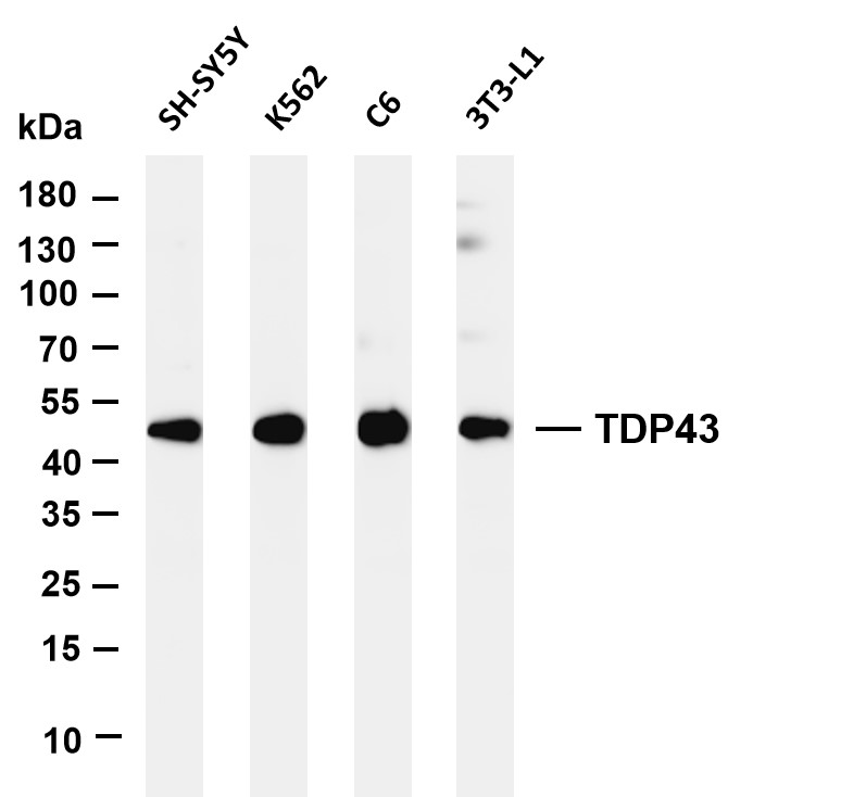
Various whole cell lysates were separated by 4-20% SDS-PAGE, and the membrane was blotted with anti-TDP43 antibody. The HRP-conjugated Goat anti-Rabbit IgG (H + L) antibody was used to detect the antibody.IHC
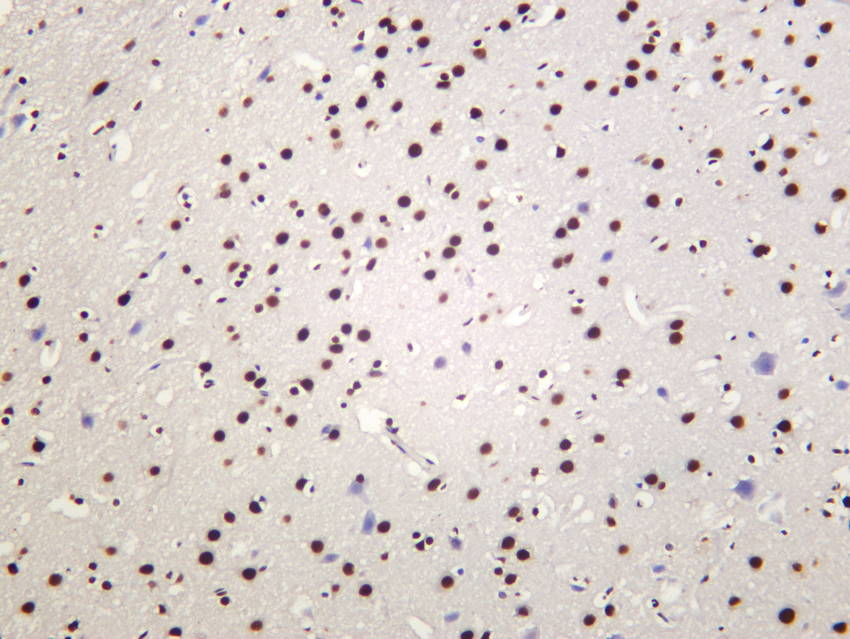
Rat brain was stained with anti-TDP43 rabbit antibody.IHC
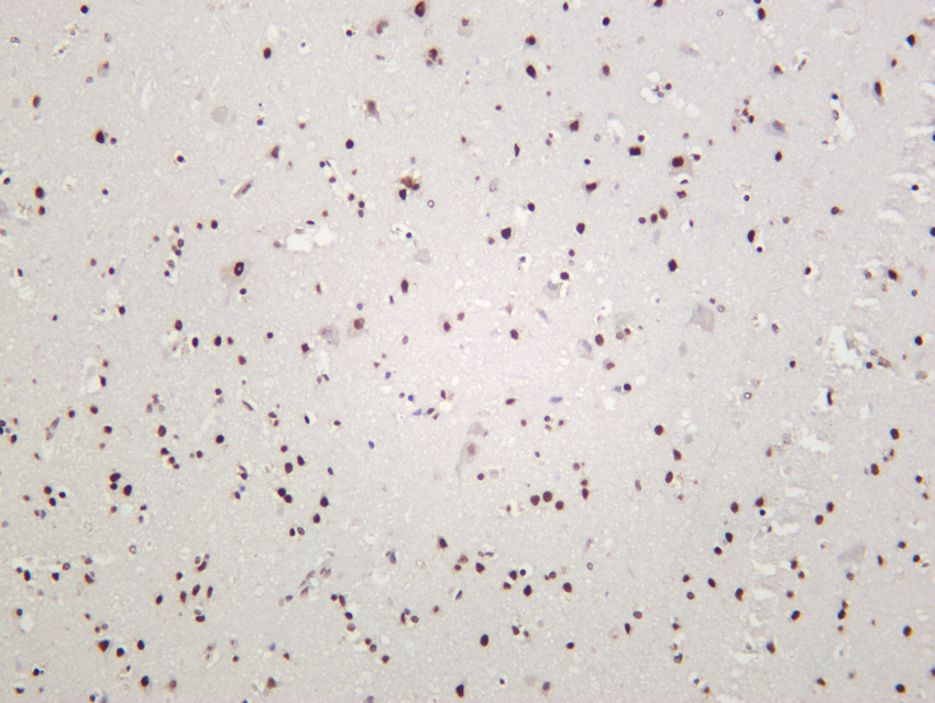
Human brain was stained with anti-TDP43 rabbit antibody.IHC
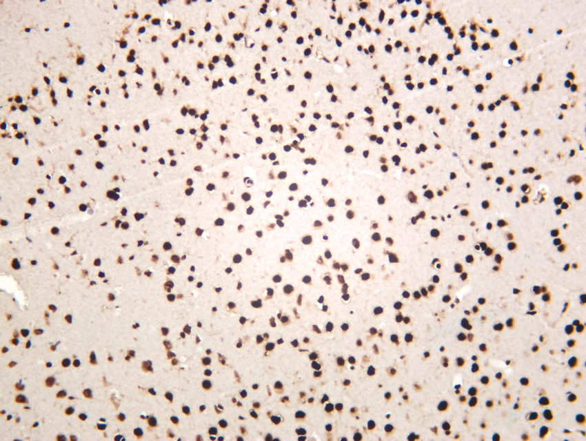
Mouse brain was stained with anti-TDP43 rabbit antibody.ICC/IF
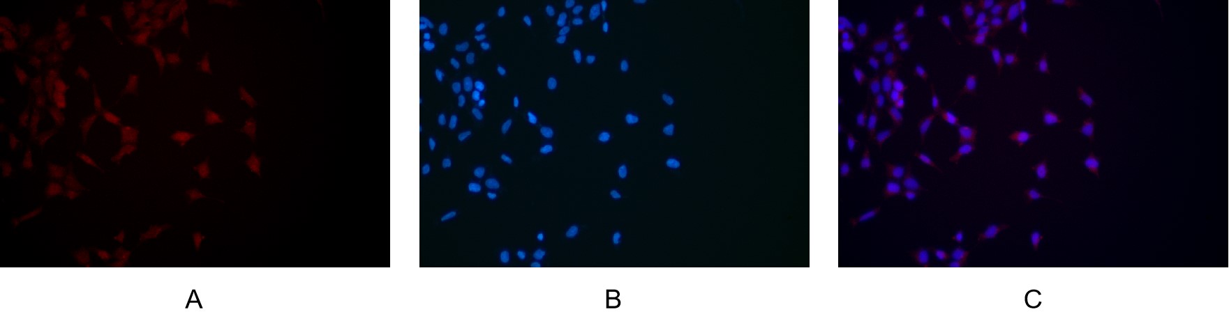
Immunofluorescence analysis of HEK293. Picture A:TDP43 antibody (red). Picture B: DAPI (blue). Picture C: Merge of A+B.| Product Name | TDP43 Rabbit mAb |
|---|---|
| Antibody Type | Primary Antibodies |
| Immunogen | The specific immunogen used to produce this antibody is proprietary information. |
| Clonality | Monoclonal |
|---|---|
| Isotype | IgG,K |
| Host Species | Rabbit |
| Tested Applications | ICC/IFIHCWB |
| WB:1:1000-1:5000 IHC:1:200-1:1000 ICC/IF:1:200-1:1000 |
|
| Species Reactivity | HumanMouseRat |
| Concentration | 1mg/ml |
| Purification | Protein A |
| Gene Symbol | TARDBP |
|---|---|
| Gene Synonyms | ALS10 TDP-43 |
| Gene Full Name | TAR DNA binding protein |
| Gene Summary | ALS10; TDP-43 Summary HIV-1, the causative agent of acquired immunodeficiency syndrome (AIDS), contains an RNA genome that produces a chromosomally integrated DNA during the replicative cycle. Activation of HIV-1 gene expression by the transactivator Tat is dependent on an RNA regulatory element (TAR) located downstream of the transcription initiation site. The protein encoded by this gene is a transcriptional repressor that binds to chromosomally integrated TAR DNA and represses HIV-1 transcription. In addition, this protein regulates alternate splicing of the CFTR gene. A similar pseudogene is present on chromosome 20. [provided by RefSeq, Jul 2008] |
| Molecular Weight(MW) | 45 kDa |
| Cellular Localization | Nucleus. |
WB
Various whole cell lysates were separated by 4-20% SDS-PAGE, and the membrane was blotted with anti-TDP43 antibody. The HRP-conjugated Goat anti-Rabbit IgG (H + L) antibody was used to detect the antibody.
IHC
Rat brain was stained with anti-TDP43 rabbit antibody.
IHC
Human brain was stained with anti-TDP43 rabbit antibody.
IHC
Mouse brain was stained with anti-TDP43 rabbit antibody.
ICC/IF
Immunofluorescence analysis of HEK293. Picture A:TDP43 antibody (red). Picture B: DAPI (blue). Picture C: Merge of A+B.| Application Notes | WB:1:1000-1:5000 IHC:1:200-1:1000 ICC/IF:1:200-1:1000 |
|---|
| Form | Liquid |
|---|---|
| Storage Instructions | -15°C to -25°C/1 year (Do not lower than -25°C). |
| Storage Buffer | PBS, 50% glycerol, 0.05% Proclin 300, 0.05%BSA. |


