IHC
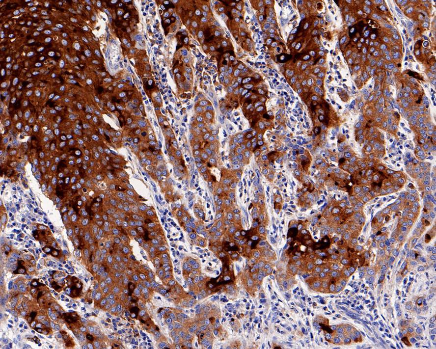
Immunohistochemical analysis of paraffin-embedded human breast carcinoma tissue with Rabbit anti-CD15 antibody at 1/1,000 dilution. The section was pre-treated using heat mediated antigen retrieval with Tris-EDTA buffer (pH 9.0) for 20 minutes. The tissues were blocked in 1% BSA for 20 minutes at room temperature, washed with ddH2O and PBS, and then probed with the primary antibody at 1/1,000 dilution for 1 hour at room temperature. The detection was performed using an HRP conjugated compact polymer system. DAB was used as the chromogen. Tissues were counterstained with hematoxylin and mounted with DPX.ICC/IF
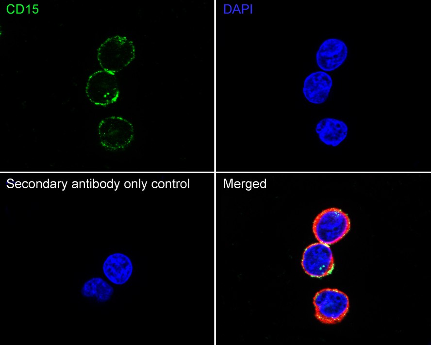
Immunocytochemistry analysis of K-562 cells labeling CD15 with Rabbit anti-CD15 antibody at 1/20 dilution. Cells were fixed in 4% paraformaldehyde for 15 minutes at room temperature, permeabilized with 0.1% Triton X-100 in PBS for 15 minutes at room temperature, then blocked with 1% BSA in 10% negative goat serum for 1 hour at room temperature. Cells were then incubated with Rabbit anti-CD15 antibody at 1/20 dilution in 1% BSA in PBST overnight at 4 ℃. Goat Anti-Rabbit IgG H&L (488) was used as the secondary antibody at 1/1,000 dilution. PBS instead of the primary antibody was used as the secondary antibody only control. Nuclear DNA was labelled in blue with DAPI. Beta tubulin (red) was stained at 1/100 dilution overnight at +4℃. Goat Anti-Mouse IgG H&L (594) was used as the secondary antibody at 1/1,000 dilution.mIHC
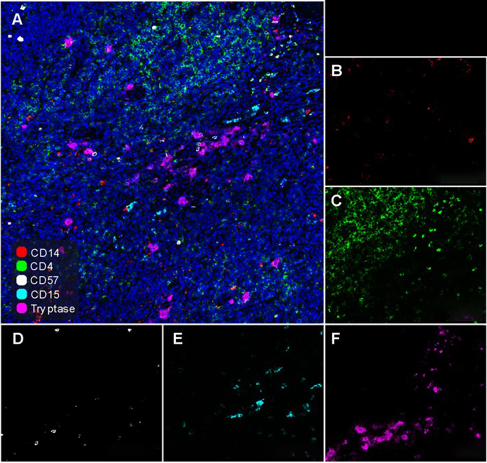
Fluorescence multiplex immunohistochemical analysis of Human tonsil (Formalin/PFA-fixed paraffin-embedded sections). Panel A: the merged image of anti-CD14 (Red), anti-CD4 (Green), anti-CD57 (White), anti-CD15 (Cyan)and anti-Tryptase (Magenta) on tonsil. Panel B: anti- CD14 stained on monocytes. Panel C: anti-CD4 stained on helper T cells and Treg cells. Panel D: anti-CD57 stained on NK cells and T cells. Panel E: CD15 stained on granulocytes and monocytes. Panel F: anti-Tryptase stained on Mast cells. Each round was followed by a separate fluorescent tyramide signal amplification system. Heat mediated antigen retrieval with Tris-EDTA buffer (pH 9.0) for 30 mins at 95℃. DAPI (blue) was used as a nuclear counter stain. Image acquisition was performed with Olympus VS200 Slide Scanner.FC
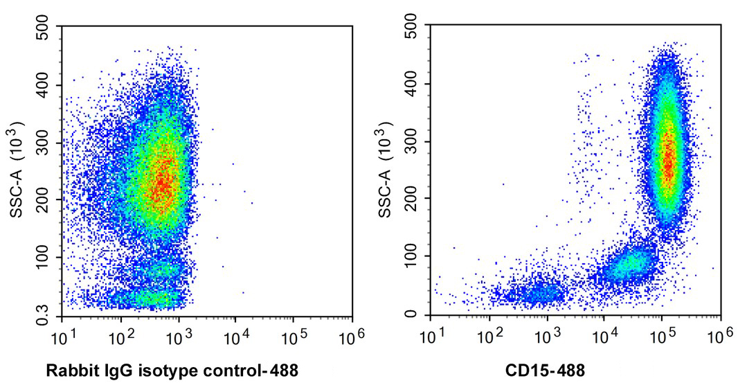
Flow cytometric analysis of human peripheral blood granulocytes labelling CD15.| Product Name | CD15 Recombinant Rabbit Monoclonal Antibody |
|---|---|
| Antibody Type | Primary Antibodies |
| Immunogen | Purified CD15 . |
| Clonality | Monoclonal |
|---|---|
| Isotype | IgG |
| Host Species | Rabbit |
| Tested Applications | FCICC/IFIHCmIHC |
| IHC:1:1000 mIHC:1:500 ICC/IF:1:20 FC:1:1000 |
|
| Species Reactivity | Human |
| Concentration | 1mg/ml |
| Purification | Protein A |
| Alternative Names | 3 fucosyl N acetyl lactosamine antibody 3-FAL antibody 3-fucosyl-N-acetyl-lactosamine antibody Leu M1 antibody Lewis X antibody LeX antibody SSEA-1 antibody stage-specific embryonic antigen 1 antibody |
|---|---|
| Molecular Weight(MW) | 59 kDa |
| Function | CD15 is a complex cluster of cell surface glycoproteins and glycolipids having a common terminal pentasaccaharide known as the Lewisx (Lex) antigen. CD15 is a haemopoietic differentiation antigen expressed on most terminally differentiated myeloid cells including granulocytes, eosinophils, mast cells, monocytes/macrophages, and Langerhans' cells. CD15 is not substantially expressed on haemopoietic progenitor cells. The positivity for CD15 is characteristic of Hodgkin’s cells in classical Hodgkin’s disease (HD). Rare cases of acute lymphoblastic leukaemia, in which myeloid antigens are often CD15 positive. Myeloid leukaemia cells express CD15 in a heterogeneous manner. CMLs are regularly CD15 positive. CD15 is expressed in a varying proportion of epithelial tumours such as adenocarcinomas (particularly from breast, lung and colon), renal cell carcinoma, apocrine carcinoma of the skin, papillary and follicular carcinoma of the thyroid, and serous carcinoma of the ovary. It is possible that sialyl-CD15 confer on the tumour cells the capacity to metastasize. Malignant mesothelioma is practically always CD15 negative (positivity has been reported in up to 6%, particularly the desmoplastic variant). In gliomas, CD15 positivity inversely correlates with the grade of malignancy. Among germ cell tumours, CD15 is detected only in mature teratoma. In haematopathology CD15 is important for the diagnosis of classical HD and characterization of acute leukaemia. CD15 may be used for histopathological grading of gliomas and differentiating between malignant gliomas and non-neoplastic glial cells (the latter usually strongly stained). Kidney and tonsil are recommended as positive and negative tissue controls for CD15. |
| Cellular Localization | Golgi apparatus, Golgi stack membrane。 |
IHC
Immunohistochemical analysis of paraffin-embedded human breast carcinoma tissue with Rabbit anti-CD15 antibody at 1/1,000 dilution. The section was pre-treated using heat mediated antigen retrieval with Tris-EDTA buffer (pH 9.0) for 20 minutes. The tissues were blocked in 1% BSA for 20 minutes at room temperature, washed with ddH2O and PBS, and then probed with the primary antibody at 1/1,000 dilution for 1 hour at room temperature. The detection was performed using an HRP conjugated compact polymer system. DAB was used as the chromogen. Tissues were counterstained with hematoxylin and mounted with DPX.
ICC/IF
Immunocytochemistry analysis of K-562 cells labeling CD15 with Rabbit anti-CD15 antibody at 1/20 dilution. Cells were fixed in 4% paraformaldehyde for 15 minutes at room temperature, permeabilized with 0.1% Triton X-100 in PBS for 15 minutes at room temperature, then blocked with 1% BSA in 10% negative goat serum for 1 hour at room temperature. Cells were then incubated with Rabbit anti-CD15 antibody at 1/20 dilution in 1% BSA in PBST overnight at 4 ℃. Goat Anti-Rabbit IgG H&L (488) was used as the secondary antibody at 1/1,000 dilution. PBS instead of the primary antibody was used as the secondary antibody only control. Nuclear DNA was labelled in blue with DAPI. Beta tubulin (red) was stained at 1/100 dilution overnight at +4℃. Goat Anti-Mouse IgG H&L (594) was used as the secondary antibody at 1/1,000 dilution.
mIHC
Fluorescence multiplex immunohistochemical analysis of Human tonsil (Formalin/PFA-fixed paraffin-embedded sections). Panel A: the merged image of anti-CD14 (Red), anti-CD4 (Green), anti-CD57 (White), anti-CD15 (Cyan)and anti-Tryptase (Magenta) on tonsil. Panel B: anti- CD14 stained on monocytes. Panel C: anti-CD4 stained on helper T cells and Treg cells. Panel D: anti-CD57 stained on NK cells and T cells. Panel E: CD15 stained on granulocytes and monocytes. Panel F: anti-Tryptase stained on Mast cells. Each round was followed by a separate fluorescent tyramide signal amplification system. Heat mediated antigen retrieval with Tris-EDTA buffer (pH 9.0) for 30 mins at 95℃. DAPI (blue) was used as a nuclear counter stain. Image acquisition was performed with Olympus VS200 Slide Scanner.
FC
Flow cytometric analysis of human peripheral blood granulocytes labelling CD15.| Application Notes | IHC:1:1000 mIHC:1:500 ICC/IF:1:20 FC:1:1000 |
|---|
| Form | Liquid |
|---|---|
| Storage Instructions | Shipped at 4℃. Store at +4℃ short term (1-2 weeks). Store at -20℃ long term. |
| Storage Buffer | PBS (pH7.4), 0.1% BSA, 40% Glycerol. Preservative: 0.05% Sodium Azide. |


