WB
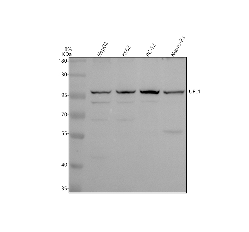
Western blot analysis of UFL1 using anti-UFL1 antibody. The sample well of each lane was loaded with 30 ug of sample under reducing conditions. After electrophoresis, proteins were transferred to a membrane. Then the membrane was incubated with rabbit anti-UFL1 antigen affinity purified polyclonal antibody at a dilution of 1:1000 and probed with a goat anti-rabbit IgG-HRP secondary antibody . The signal is developed using ECL Plus Western Blotting Substrate.IHC
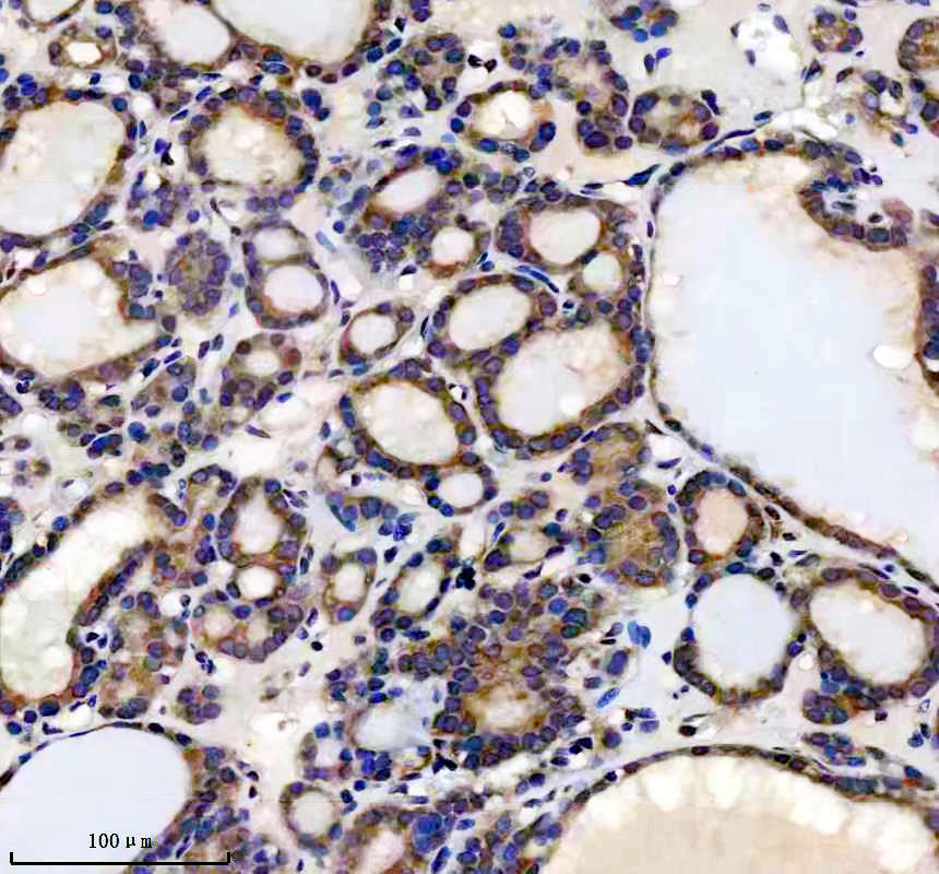
IHC analysis of UFL1 using anti-UFL1 antibody. UFL1 was detected in a paraffin-embedded section of human thyroid cancer tissue. The tissue section was incubated with rabbit anti-UFL1 Antibody at a dilution of 1:200 and developed using HRP Conjugated Rabbit IgG Super Vision Assay Kit with DAB as the chromogen.| Product Name | Rabbit polyclonal antibody to UFL1 |
|---|---|
| Antibody Type | Primary Antibodies |
| Immunogen | E.coli-derived human UFL1 recombinant protein (Position: 3-347). |
| Clonality | Polyclonal |
|---|---|
| Isotype | IgG |
| Host Species | Rabbit |
| Tested Applications | IHCWB |
| WB:1:500-1:2000 IHC:1:50-1:400 |
|
| Species Reactivity | HumanMouseRat |
| Concentration | 0.5mg/ml |
| Purification | Affinity purified |
| Gene Symbol | UFL1 |
|---|---|
| Gene Synonyms | NLBP RCAD Maxer KIAA0776 |
| Gene Full Name | UFM1 specific ligase 1 |
| Gene Summary | Enables UFM1 ligase activity and protein kinase binding activity. Involved in several processes, including positive regulation of reticulophagy; regulation of intracellular signal transduction; and regulation of protein metabolic process. Acts upstream of or within several processes, including positive regulation of cell population proliferation; regulation of proteasomal ubiquitin-dependent protein catabolic process; and response to endoplasmic reticulum stress. Located in cytoplasm; nucleus; and site of double-strand break. Part of protein-containing complex. Is active in endoplasmic reticulum membrane and mitochondrial outer membrane. [provided by Alliance of Genome Resources, Jun 2026] |
| Alternative Names | UFL1 KIAA0776 MAXER NLBP RCAD E3 UFM1-protein ligase 1 EC 2.3.2.- E3 UFM1-protein transferase 1 Multiple alpha-helix protein located at ER Novel LZAP-binding protein Regulator of C53/LZAP and DDRGK1 |
| Molecular Weight(MW) | 90 kDa(Observed band size:100 kDa) |
| Cellular Localization | Chromosome,Cytoplasm,Endoplasmic reticulum,Membrane,Nucleus. |
WB
Western blot analysis of UFL1 using anti-UFL1 antibody. The sample well of each lane was loaded with 30 ug of sample under reducing conditions. After electrophoresis, proteins were transferred to a membrane. Then the membrane was incubated with rabbit anti-UFL1 antigen affinity purified polyclonal antibody at a dilution of 1:1000 and probed with a goat anti-rabbit IgG-HRP secondary antibody . The signal is developed using ECL Plus Western Blotting Substrate.
IHC
IHC analysis of UFL1 using anti-UFL1 antibody. UFL1 was detected in a paraffin-embedded section of human thyroid cancer tissue. The tissue section was incubated with rabbit anti-UFL1 Antibody at a dilution of 1:200 and developed using HRP Conjugated Rabbit IgG Super Vision Assay Kit with DAB as the chromogen.| Application Notes | WB:1:500-1:2000 IHC:1:50-1:400 |
|---|
| Form | Liquid |
|---|---|
| Storage Instructions | 12 months from date of receipt,-20℃ as supplied. |
| Storage Buffer | 500 ug/ml antibody with PBS, 0.02% NaN3, 1 mg/ml BSA and 50% glycerol. |


