WB
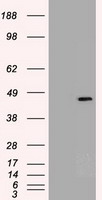
HEK293T cells were transfected with the pCMV6-ENTRY control (Left lane) or pCMV6-ENTRY SOX17 (Right lane) cDNA for 48 hrs and lysed. Equivalent amounts of cell lysates (5 ug per lane) were separated by SDS-PAGE and immunoblotted with anti-SOX17.IHC
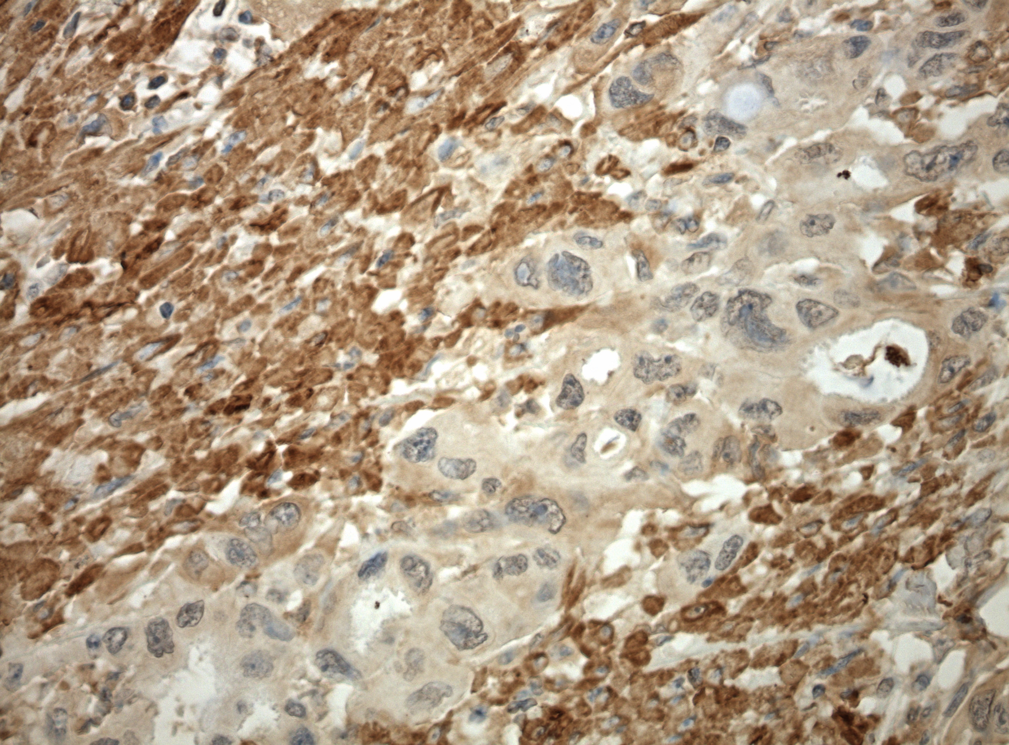
Immunohistochemical staining of paraffin-embedded pancreatic carcinoma using anti-SOX17 antibody clone mouse monoclonal antibody. Protocol used HIER TEE pH9.0 and anti-SOX17 at 1:100 dilution. Detection was done with Polink1 Broad Mouse and Rabbit C/N D11-18 with DAB Kit. Image 40x magnification.IHC
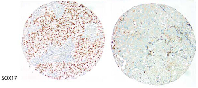
Figure from citation: Corresponding conventional and immunhistochemical stains of human seminoma and embryonal carcinoma by using anti-SOX17 antibody. Seminoma: positivity for SOX17 (Left); Embryonal carcinoma: negative staining for SOX17 (Right). Dilution: 1:2000 View Citation. Heat-induced epitope retrieval by EDTA solution buffer pH 8.0 at 120°C for 3 min.ICC/IF
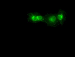
Anti-SOX17 mouse monoclonal antibody immunofluorescent staining of COS7 cells transiently transfected by pCMV6-ENTRY SOX17.| Product Name | SOX17 Mouse Monoclonal Antibody |
|---|---|
| Antibody Type | Primary Antibodies |
| Immunogen | Human recombinant protein fragment corresponding to amino acids 177-414 of human SOX17 (NP_071899) produced in E.coli. |
| Clonality | Monoclonal |
|---|---|
| Isotype | IgG2b |
| Host Species | Mouse |
| Tested Applications | ICC/IFIHCWB |
| WB:1:1000~1:2000 IHC:1:50 ICC/IF:1:50~1:100 |
|
| Species Reactivity | DogHumanMouseRat |
| Concentration | 1mg/ml |
| Purification | Protein A/G |
| Gene Symbol | SOX17 |
|---|---|
| Gene Synonyms | PPH7 VUR3 |
| Gene Full Name | SRY-box transcription factor 17 |
| Gene Summary | This gene encodes a member of the SOX (SRY-related HMG-box) family of transcription factors involved in the regulation of embryonic development and in the determination of the cell fate. The encoded protein may act as a transcriptional regulator after forming a protein complex with other proteins. [provided by RefSeq, Jul 2008] |
| Alternative Names | VUR3 |
| Molecular Weight(MW) | 43.9 kDa |
| Cellular Localization | Nucleus. |
WB
HEK293T cells were transfected with the pCMV6-ENTRY control (Left lane) or pCMV6-ENTRY SOX17 (Right lane) cDNA for 48 hrs and lysed. Equivalent amounts of cell lysates (5 ug per lane) were separated by SDS-PAGE and immunoblotted with anti-SOX17.
IHC
Immunohistochemical staining of paraffin-embedded pancreatic carcinoma using anti-SOX17 antibody clone mouse monoclonal antibody. Protocol used HIER TEE pH9.0 and anti-SOX17 at 1:100 dilution. Detection was done with Polink1 Broad Mouse and Rabbit C/N D11-18 with DAB Kit. Image 40x magnification.
IHC
Figure from citation: Corresponding conventional and immunhistochemical stains of human seminoma and embryonal carcinoma by using anti-SOX17 antibody. Seminoma: positivity for SOX17 (Left); Embryonal carcinoma: negative staining for SOX17 (Right). Dilution: 1:2000 View Citation. Heat-induced epitope retrieval by EDTA solution buffer pH 8.0 at 120°C for 3 min.
ICC/IF
Anti-SOX17 mouse monoclonal antibody immunofluorescent staining of COS7 cells transiently transfected by pCMV6-ENTRY SOX17.| Application Notes | WB:1:1000~1:2000 IHC:1:50 ICC/IF:1:50~1:100 |
|---|
| Form | Liquid |
|---|---|
| Storage Instructions | Store at -20°C as received. |
| Storage Buffer | PBS (pH 7.3) containing 1% BSA, 50% glycerol and 0.02% sodium azide. |


