WB
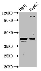
Western Blot. Positive WB detected in: U251 whole cell lysate, HepG2 whole cell lysate. All lanes: C1GALT1 antibody at 4.4µg/ml. Secondary Goat polyclonal to rabbit IgG at 1/50000 dilution.IHC
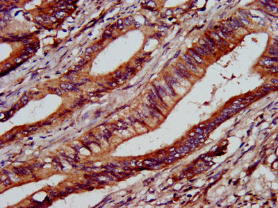
IHC image of C1GALT1 Antibody diluted at 1:1000 and staining in paraffin-embedded human colon cancer performed on a Leica BondTM system. After dewaxing and hydration, antigen retrieval was mediated by high pressure in a citrate buffer (pH 6.0). Section was blocked with 10% normal goat serum 30min at RT. Then primary antibody (1% BSA) was incubated at 4°C overnight. The primary is detected by a biotinylated secondary antibody and visualized using an HRP conjugated SP system.ICC/IF
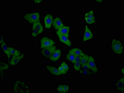
Immunofluorescence staining of HepG2 cells with C1GALT1 Antibody at 1:333, counter-stained with DAPI. The cells were fixed in 4% formaldehyde, permeabilized using 0.2% Triton X-100 and blocked in 10% normal Goat Serum. The cells were then incubated with the antibody overnight at 4°C. The secondary antibody was Alexa Fluor 488-congugated AffiniPure Goat Anti-Rabbit IgG(H+L).| Product Name | Rabbit anti-C1GALT1 Polyclonal antibody |
|---|---|
| Antibody Type | Primary Antibodies |
| Immunogen | Recombinant Human Glycoprotein-N-acetylgalactosamine 3-beta-galactosyltransferase 1 protein (34-143AA). |
| Clonality | Polyclonal |
|---|---|
| Isotype | IgG |
| Host Species | Rabbit |
| Tested Applications | ICC/IFIHCWB |
| WB:1:500-1:5000 IHC:1:500-1:1000 ICC/IF:1:200-1:500 |
|
| Species Reactivity | Human |
| Concentration | 1mg/ml |
| Purification | Protein G |
| Gene Symbol | C1GALT1 |
|---|---|
| Gene Synonyms | C1GALT T-synthase |
| Gene Full Name | core 1 synthase, glycoprotein-N-acetylgalactosamine 3-beta-galactosyltransferase 1 |
| Gene Summary | The protein encoded by this gene generates the common core 1 O-glycan structure, Gal-beta-1-3GalNAc-R, by the transfer of Gal from UDP-Gal to GalNAc-alpha-1-R. Core 1 is a precursor for many extended mucin-type O-glycans on cell surface and secreted glycoproteins. Studies in mice suggest that this gene plays a key role in thrombopoiesis and kidney homeostasis.[provided by RefSeq, Sep 2010] |
| Alternative Names | C1GALT1Glycoprotein-N-acetylgalactosamine 3-beta-galactosyltransferase 1 antibody EC 2.4.1.122 antibody B3Gal-T8 antibody Core 1 O-glycan T-synthase antibody Core 1 UDP-galactose:N-acetylgalactosamine-alpha-R beta 1 3-galactosyltransferase 1 antibody Beta-1 3-galactosyltransferase antibody Core 1 beta1 3-galactosyltransferase 1 antibody C1GalT1 antibody Core 1 beta3-Gal-T1 antibody |
| Molecular Weight(MW) | 43, 36 kDa(Observed band size: 43 kDa) |
| Cellular Localization | Membrane. |
WB
Western Blot. Positive WB detected in: U251 whole cell lysate, HepG2 whole cell lysate. All lanes: C1GALT1 antibody at 4.4µg/ml. Secondary Goat polyclonal to rabbit IgG at 1/50000 dilution.
IHC
IHC image of C1GALT1 Antibody diluted at 1:1000 and staining in paraffin-embedded human colon cancer performed on a Leica BondTM system. After dewaxing and hydration, antigen retrieval was mediated by high pressure in a citrate buffer (pH 6.0). Section was blocked with 10% normal goat serum 30min at RT. Then primary antibody (1% BSA) was incubated at 4°C overnight. The primary is detected by a biotinylated secondary antibody and visualized using an HRP conjugated SP system.
ICC/IF
Immunofluorescence staining of HepG2 cells with C1GALT1 Antibody at 1:333, counter-stained with DAPI. The cells were fixed in 4% formaldehyde, permeabilized using 0.2% Triton X-100 and blocked in 10% normal Goat Serum. The cells were then incubated with the antibody overnight at 4°C. The secondary antibody was Alexa Fluor 488-congugated AffiniPure Goat Anti-Rabbit IgG(H+L).| Application Notes | WB:1:500-1:5000 IHC:1:500-1:1000 ICC/IF:1:200-1:500 |
|---|
| Form | Liquid |
|---|---|
| Storage Instructions | Upon receipt, store at -20°C or -80°C. Avoid repeated freeze. |
| Storage Buffer | Preservative: 0.03% Proclin 300; Constituents: 50% Glycerol, 0.01M PBS, pH 7.4. |


