WB
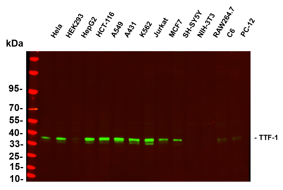
Various whole cell lysates were separated by 4-20% SDS-PAGE, and the primary antibody was used at 4℃ over night with a 1:5000 dilution . The Dylight 800-conjugated Goat anti-Rabbit antibody was used to detect the antibody.WB
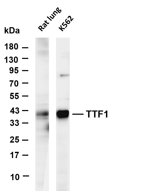
Various whole cell lysates were separated by 4-20% SDS-PAGE, and the membrane was blotted with anti-TTF1 antibody. The HRP-conjugated Goat anti-Rabbit IgG (H + L) antibody was used to detect the antibody.IHC
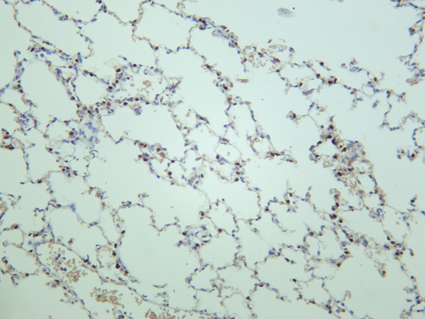
Rat lung was stained with anti-TTF1 rabbit antibody.IHC
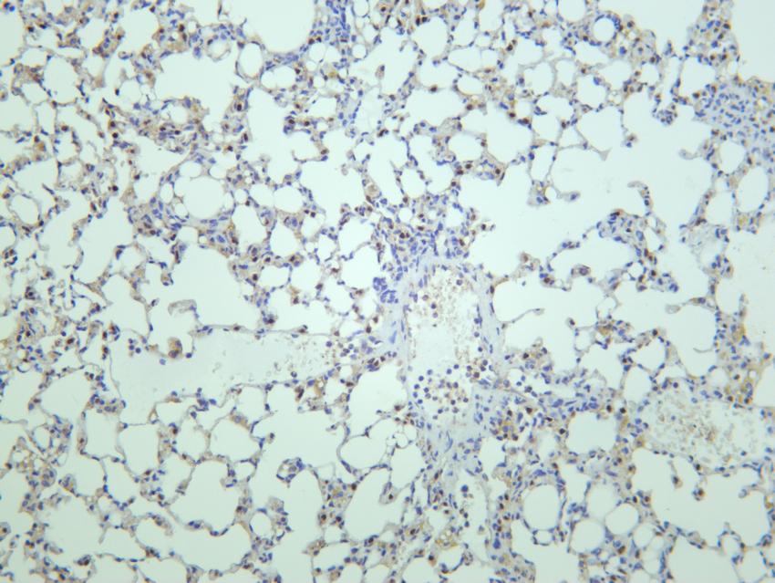
Mouse lung was stained with anti-TTF1 rabbit antibody.IHC
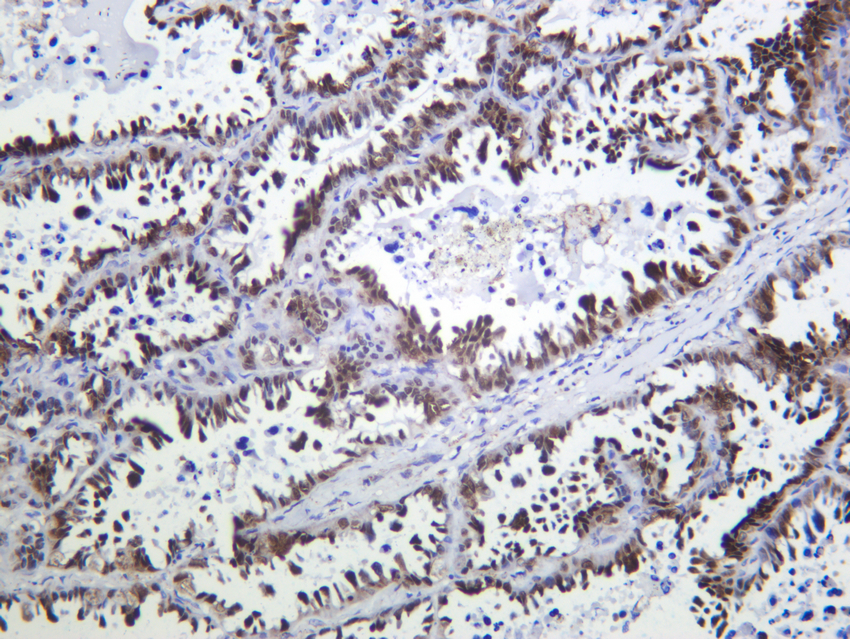
Human lung adenocarcinoma was stained with anti-TTF-1 Rabbit antibody.IF-P
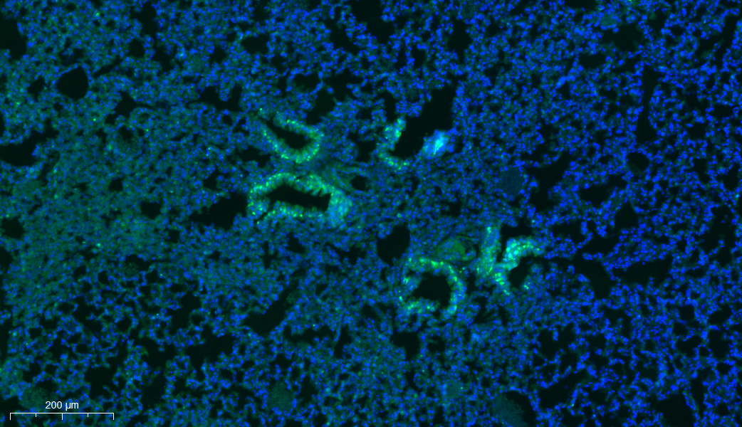
Immunofluorescence multiplex immunohistochemical analysis of Rat lung (formalin-fixed paraffin-embedded section). using 488(Green) fluorescent tyramide signal amplification system. Antigen retrieval (pH 9.0, 20 minutes) was used before antibody incubation. DAPI (dark blue) was used as a nuclear counter stain.| Product Name | TTF-1 Rabbit mAb |
|---|---|
| Antibody Type | Primary Antibodies |
| Immunogen | The specific immunogen used to produce this antibody is proprietary information. |
| Clonality | Monoclonal |
|---|---|
| Isotype | IgG,K |
| Host Species | Rabbit |
| Tested Applications | IF-PIHCWB |
| WB:1:2000-1:10000 IHC:1:200-1:1000 IF-P:1:200-1:1000 |
|
| Species Reactivity | HumanMouseRat |
| Concentration | 1mg/ml |
| Purification | Protein A |
| Gene Symbol | NKX2-1 |
|---|---|
| Gene Synonyms | BCH BHC NK-2 TEBP TTF1 NKX2A NMTC1 T/EBP TITF1 TTF-1 NKX2.1 |
| Gene Full Name | NK2 homeobox 1 |
| Gene Summary | This gene encodes a protein initially identified as a thyroid-specific transcription factor. The encoded protein binds to the thyroglobulin promoter and regulates the expression of thyroid-specific genes but has also been shown to regulate the expression of genes involved in morphogenesis. Mutations and deletions in this gene are associated with benign hereditary chorea, choreoathetosis, congenital hypothyroidism, and neonatal respiratory distress, and may be associated with thyroid cancer. Multiple transcript variants encoding different isoforms have been found for this gene. This gene shares the symbol/alias 'TTF1' with another gene, transcription termination factor 1, which plays a role in ribosomal gene transcription. [provided by RefSeq, Feb 2014] |
| Alternative Names | NKX2-1 NKX2A TITF1 TTF1 Homeobox protein Nkx-2.1 Homeobox protein NK-2 homolog A Thyroid nuclear factor 1 Thyroid transcription factor 1 TTF-1 |
| Molecular Weight(MW) | 40 kDa |
| Cellular Localization | Nuclear. |
WB
Various whole cell lysates were separated by 4-20% SDS-PAGE, and the primary antibody was used at 4℃ over night with a 1:5000 dilution . The Dylight 800-conjugated Goat anti-Rabbit antibody was used to detect the antibody.
WB
Various whole cell lysates were separated by 4-20% SDS-PAGE, and the membrane was blotted with anti-TTF1 antibody. The HRP-conjugated Goat anti-Rabbit IgG (H + L) antibody was used to detect the antibody.
IHC
Rat lung was stained with anti-TTF1 rabbit antibody.
IHC
Mouse lung was stained with anti-TTF1 rabbit antibody.
IHC
Human lung adenocarcinoma was stained with anti-TTF-1 Rabbit antibody.
IF-P
Immunofluorescence multiplex immunohistochemical analysis of Rat lung (formalin-fixed paraffin-embedded section). using 488(Green) fluorescent tyramide signal amplification system. Antigen retrieval (pH 9.0, 20 minutes) was used before antibody incubation. DAPI (dark blue) was used as a nuclear counter stain.| Application Notes | WB:1:2000-1:10000 IHC:1:200-1:1000 IF-P:1:200-1:1000 |
|---|
| Form | Liquid |
|---|---|
| Storage Instructions | -15°C to -25°C/1 year (Do not lower than -25°C). |
| Storage Buffer | PBS, 50% glycerol, 0.05% Proclin 300, 0.05%BSA. |


