WB
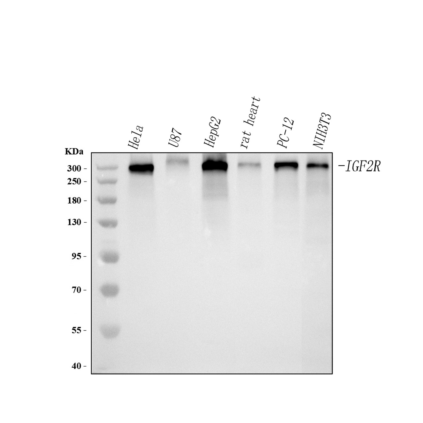
Western blot analysis of anti-IGF2R antibody. The sample well of each lane was loaded with 30 ug of sample under reducing conditions. After electrophoresis, proteins were transferred to a membrane. Then the membrane was incubated with rabbit anti-IGF2R antigen affinity purified polyclonal antibody and probed with a goat anti-rabbit IgG-HRP secondary antibody. The signal is developed using ECL Plus Western Blotting Substrate.IHC
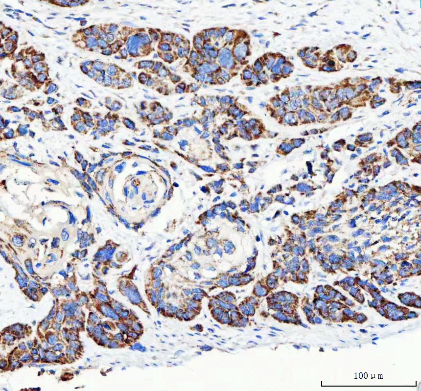
IHC analysis of IGF2R using anti-IGF2R antibody. IGF2R was detected in a paraffin-embedded section of human esophageal squamous carcinoma tissue. The tissue section was developed using HRP Conjugated Rabbit IgG Super Vision Assay Kit with DAB as the chromogen.ICC/IF
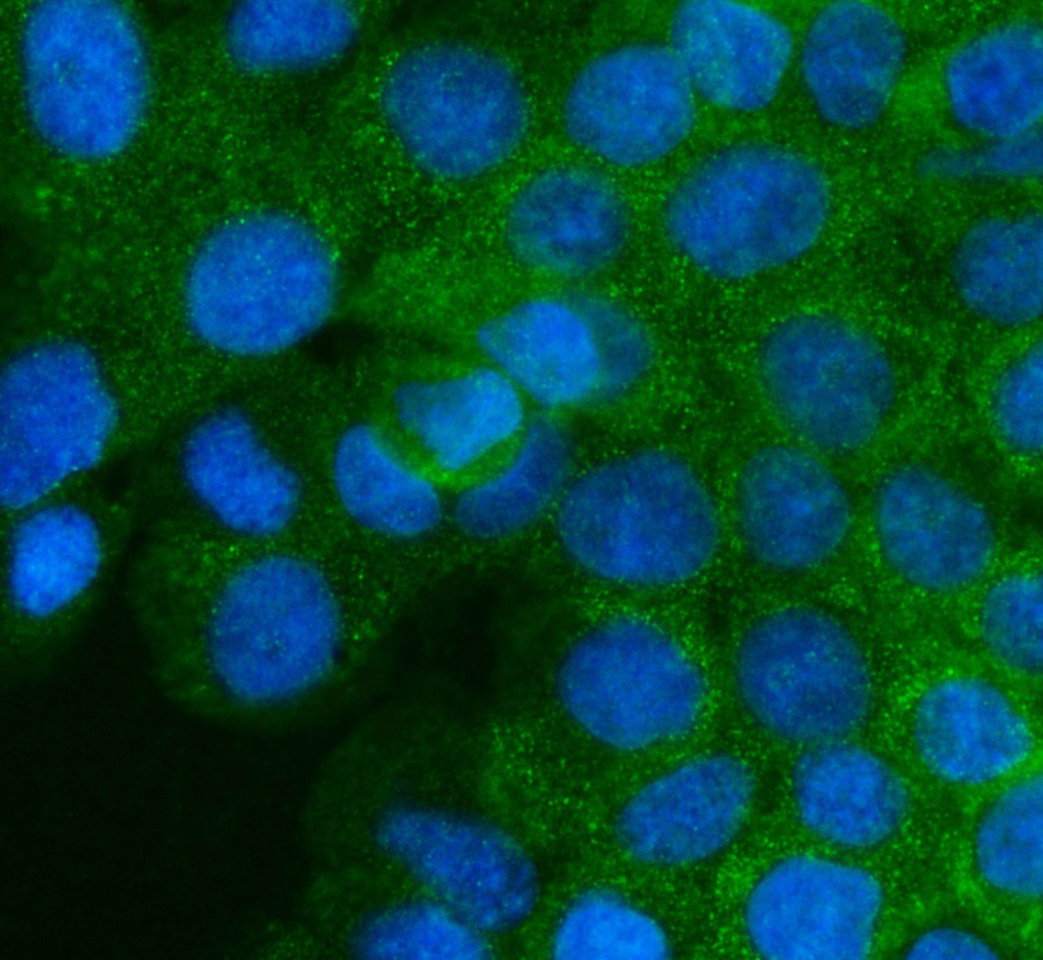
ICC/IF analysis of IGF2R using anti-IGF2R antibody. IGF2R was detected in an immunocytochemical section of A431 cells. Fluoro488 Conjugated Goat Anti-Rabbit IgG (Green) was used as secondary antibody. The section was counterstained with DAPI (Blue).FC
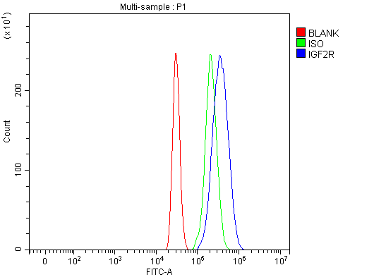
Flow Cytometry analysis of HepG2 cells using anti-IGF2R antibody. Overlay histogram showing HepG2 cells stained with Anti-IGF2R Antibody (Blue line). To facilitate intracellular staining, cells were fixed with 4% paraformaldehyde and permeabilized with permeabilization buffer. The cells were blocked with 10% normal goat serum. And then incubated with rabbit anti-IGF2R Antibody (1:100). Fluoro488 conjugated goat anti-rabbit IgG (1:100) was used as secondary antibody. Isotype control antibody (Green line) was rabbit IgG (1:100) used under the same conditions. Unlabelled sample (Red line) was also used as a control.| Product Name | Rabbit polyclonal antibody to IGF2R |
|---|---|
| Antibody Type | Primary Antibodies |
| Immunogen | E. coli-derived human IGF2R recombinant protein (Position: F424-R529). |
| Clonality | Polyclonal |
|---|---|
| Isotype | IgG |
| Host Species | Rabbit |
| Tested Applications | FCICC/IFIHCWB |
| WB:1:500-1:2000 IHC:1:50-1:400 ICC/IF:1:50-1:400 FC:1:50-1:200 |
|
| Species Reactivity | HumanMouseRat |
| Concentration | 0.5mg/ml |
| Purification | Affinity purified |
| Gene Symbol | IGF2R |
|---|---|
| Gene Synonyms | MPR1 MPRI CD222 CIMPR M6P-R MPR300 CI-M6PR MPR 300 M6P/IGF2R |
| Gene Full Name | insulin like growth factor 2 receptor |
| Gene Summary | This gene encodes a receptor for both insulin-like growth factor 2 and mannose 6-phosphate. The binding sites for each ligand are located on different segments of the protein. This receptor has various functions, including in the intracellular trafficking of lysosomal enzymes, the activation of transforming growth factor beta, and the degradation of insulin-like growth factor 2. Mutation or loss of heterozygosity of this gene has been association with risk of hepatocellular carcinoma. The orthologous mouse gene is imprinted and shows exclusive expression from the maternal allele; however, imprinting of the human gene may be polymorphic, as only a minority of individuals showed biased expression from the maternal allele (PMID:8267611). [provided by RefSeq, Nov 2015] |
| Alternative Names | CD222 CI Man 6 P receptor CI MPR CIMPR IGF II receptor IGF2R IGF2R-Specific Lysosome Marker M6P R M6P/IGF2 receptor M6P/IGF2R M6PR MPR 300 MPR1 MPRI |
| Molecular Weight(MW) | 274 kDa |
| Cellular Localization | Endosome,Golgi apparatus,Membrane. |
WB
Western blot analysis of anti-IGF2R antibody. The sample well of each lane was loaded with 30 ug of sample under reducing conditions. After electrophoresis, proteins were transferred to a membrane. Then the membrane was incubated with rabbit anti-IGF2R antigen affinity purified polyclonal antibody and probed with a goat anti-rabbit IgG-HRP secondary antibody. The signal is developed using ECL Plus Western Blotting Substrate.
IHC
IHC analysis of IGF2R using anti-IGF2R antibody. IGF2R was detected in a paraffin-embedded section of human esophageal squamous carcinoma tissue. The tissue section was developed using HRP Conjugated Rabbit IgG Super Vision Assay Kit with DAB as the chromogen.
ICC/IF
ICC/IF analysis of IGF2R using anti-IGF2R antibody. IGF2R was detected in an immunocytochemical section of A431 cells. Fluoro488 Conjugated Goat Anti-Rabbit IgG (Green) was used as secondary antibody. The section was counterstained with DAPI (Blue).
FC
Flow Cytometry analysis of HepG2 cells using anti-IGF2R antibody. Overlay histogram showing HepG2 cells stained with Anti-IGF2R Antibody (Blue line). To facilitate intracellular staining, cells were fixed with 4% paraformaldehyde and permeabilized with permeabilization buffer. The cells were blocked with 10% normal goat serum. And then incubated with rabbit anti-IGF2R Antibody (1:100). Fluoro488 conjugated goat anti-rabbit IgG (1:100) was used as secondary antibody. Isotype control antibody (Green line) was rabbit IgG (1:100) used under the same conditions. Unlabelled sample (Red line) was also used as a control.| Application Notes | WB:1:500-1:2000 IHC:1:50-1:400 ICC/IF:1:50-1:400 FC:1:50-1:200 |
|---|
| Form | Liquid |
|---|---|
| Storage Instructions | 12 months from date of receipt,-20℃ as supplied. |
| Storage Buffer | 500 ug/ml antibody with PBS, 0.02% NaN3, 1 mg/ml BSA and 50% glycerol. |


