WB
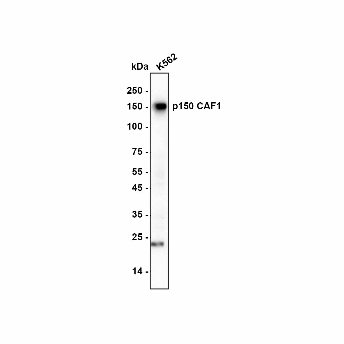
Western blot analysis of p150 CAF1 on K562 cell lysate lysate with Rabbit anti-p150 CAF1 antibody at 1/1,000 dilution. Lysates/proteins at 15 µg/Lane. Exposure time: 20 seconds; Blocking: 5% NFDM/TBST, 1 hour at room temperature. Primary antibody: p150 CAF1 Recombinant Rabbit Monoclonal Antibody, 1/1,000 in primary antibody dilution buffer, overnight at 4 ℃. Secondary antibody: Goat anti-Rabbit IgG-HRP, 1/50,000 in 5% NFDM/TBST, 1 hour at room temperature.IHC
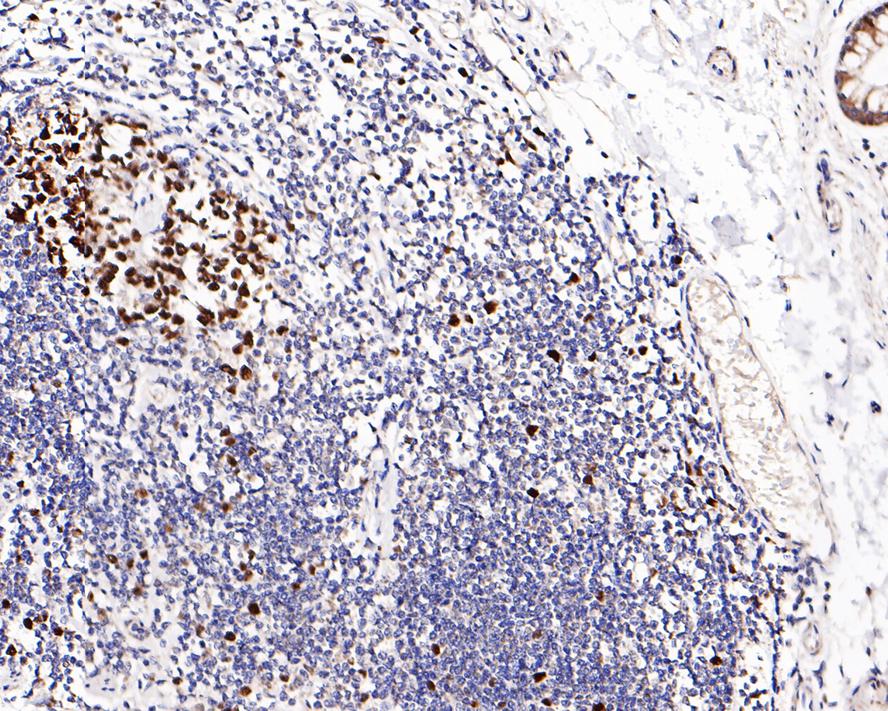
Immunohistochemical analysis of paraffin-embedded human colon tissue with Rabbit anti-p150 CAF1 antibody at 1/200 dilution. The section was pre-treated using heat mediated antigen retrieval with sodium citrate buffer (pH 6.0) (high pressure) for 2 minutes. The tissues were blocked in 1% BSA for 20 minutes at room temperature, washed with ddH2O and PBS, and then probed with the primary antibody at 1/200 dilution for 1 hour at room temperature. The detection was performed using an HRP conjugated compact polymer system. DAB was used as the chromogen. Tissues were counterstained with hematoxylin and mounted with DPX.ICC/IF
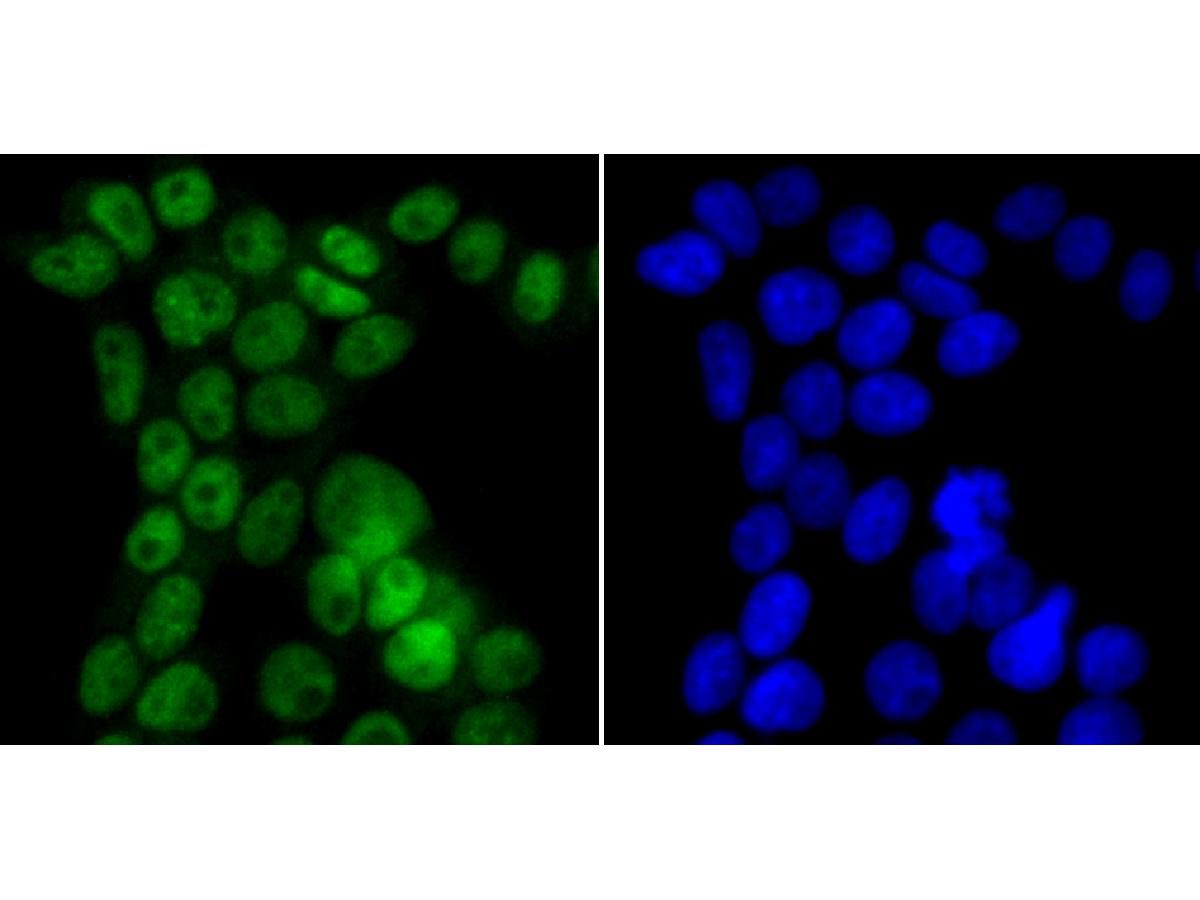
ICC staining p150 CAF1 in 293T cells (green). The nuclear counter stain is DAPI (blue). Cells were fixed in paraformaldehyde, permeabilised with 0.25% Triton X100/PBS.| Product Name | p150 CAF1 Recombinant Rabbit Monoclonal Antibody |
|---|---|
| Antibody Type | Primary Antibodies |
| Immunogen | Recombinant protein within Human p150 CAF1 aa 81-195 / 956. |
| Clonality | Monoclonal |
|---|---|
| Isotype | IgG |
| Host Species | Rabbit |
| Tested Applications | ICC/IFIHCWB |
| WB:1:500-1:2000 IHC:1:50-1:200 ICC/IF:1:50-1:200 |
|
| Species Reactivity | Human |
| Concentration | 1mg/ml |
| Purification | Protein A |
| Gene Symbol | CHAF1A |
|---|---|
| Gene Synonyms | CAF1 P150 CAF-1 CAF1B CAF1P150 |
| Gene Full Name | chromatin assembly factor 1 subunit A |
| Gene Summary | Chromatin assembly factor I (CAF1) is a nuclear complex consisting of p50, p60 (CHAF1B; MIM 601245), and p150 (CHAF1A) subunits that assembles histone octamers onto replicating DNA in vitro (Kaufman et al., 1995 [PubMed 7600578]).[supplied by OMIM, Mar 2008] |
| Alternative Names | CAF 1 antibody CAF 1 subunit A antibody CAF antibody CAF I 150 kDa subunit antibody CAF I p150 antibody CAF-1 subunit A antibody CAF-I 150 kDa subunit antibody CAF-I p150 antibody CAF1 antibody CAF1A_HUMAN antibody CAF1B antibody CAF1P150 antibody CHAF1A antibody Chromatin assembly factor 1 subunit A antibody Chromatin Assembly Factor 1 Subunit A p150 antibody Chromatin assembly factor I (150 kDa) antibody Chromatin assembly factor I 150 kDa antibody Chromatin assembly factor I p150 subunit antibody DCAF1 antibody hp150 antibody MGC71229 antibody P150 antibody |
| Molecular Weight(MW) | 107 kDa(Observed band size: 150 kDa) |
| Cellular Localization | Nucleus. |
WB
Western blot analysis of p150 CAF1 on K562 cell lysate lysate with Rabbit anti-p150 CAF1 antibody at 1/1,000 dilution. Lysates/proteins at 15 µg/Lane. Exposure time: 20 seconds; Blocking: 5% NFDM/TBST, 1 hour at room temperature. Primary antibody: p150 CAF1 Recombinant Rabbit Monoclonal Antibody, 1/1,000 in primary antibody dilution buffer, overnight at 4 ℃. Secondary antibody: Goat anti-Rabbit IgG-HRP, 1/50,000 in 5% NFDM/TBST, 1 hour at room temperature.
IHC
Immunohistochemical analysis of paraffin-embedded human colon tissue with Rabbit anti-p150 CAF1 antibody at 1/200 dilution. The section was pre-treated using heat mediated antigen retrieval with sodium citrate buffer (pH 6.0) (high pressure) for 2 minutes. The tissues were blocked in 1% BSA for 20 minutes at room temperature, washed with ddH2O and PBS, and then probed with the primary antibody at 1/200 dilution for 1 hour at room temperature. The detection was performed using an HRP conjugated compact polymer system. DAB was used as the chromogen. Tissues were counterstained with hematoxylin and mounted with DPX.
ICC/IF
ICC staining p150 CAF1 in 293T cells (green). The nuclear counter stain is DAPI (blue). Cells were fixed in paraformaldehyde, permeabilised with 0.25% Triton X100/PBS.| Application Notes | WB:1:500-1:2000 IHC:1:50-1:200 ICC/IF:1:50-1:200 |
|---|
| Form | Liquid |
|---|---|
| Storage Instructions | Shipped at 4℃. Store at +4℃ short term (1-2 weeks). Store at -20℃ long term. |
| Storage Buffer | 1*TBS (pH7.4), 0.05% BSA, 40% Glycerol. Preservative: 0.05% Sodium Azide. |


