WB
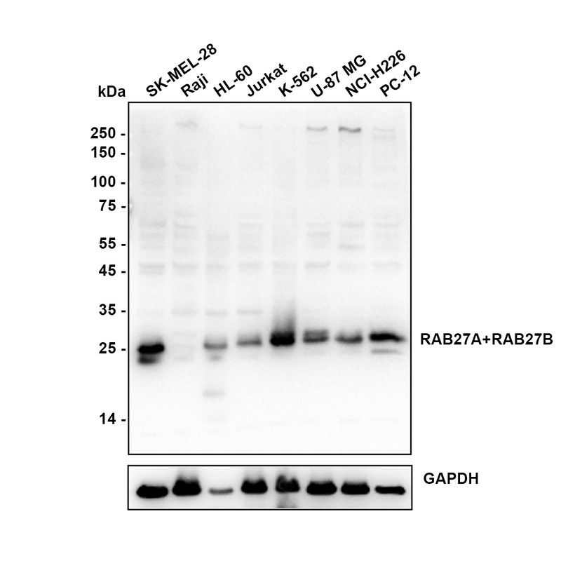
Western blot analysis of RAB27A+RAB27B on different lysates with Rabbit anti-RAB27A+RAB27B antibody at 1/5,000 dilution. Lysates/proteins at 15 µg/Lane. Exposure time: 10 seconds; Negative control: Raji. Blocking: 5% NFDM/TBST, 1 hour at room temperature. Primary antibody: RAB27A+RAB27B Recombinant Rabbit Monoclonal Antibody, 1/5,000 in primary antibody dilution buffer, overnight at 4 ℃. Secondary antibody: Goat anti-Rabbit IgG-HRP, 1/50,000 in 5% NFDM/TBST, 1 hour at room temperature.FC
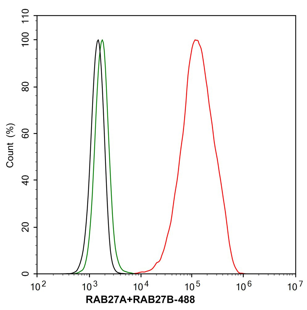
Application: Flow Cytometry (Intra). Species: Rat. Sample: PC-12 (Rat pheochromocytoma cells (undifferentiated)). Fixation: 4% Paraformaldehyde, 15 minutes at room temperature. Permeabilization: 1x Permeabilization Buffer (eBioscience), 15 minutes at room temperature. Blocking: 1% BSA + 10% normal goat serum, 15 minutes at room temperature. Antibody dilution buffer: 1x PBS. Primary antibody: RAB27A+RAB27B Recombinant Rabbit Monoclonal Antibody(1/1,000, Red) compared with Rabbit IgG Isotype Control (Green), 15 minutes at room temperature. Secondary antibody: Goat Anti-Rabbit IgG (488), 15 minutes at room temperature.IP
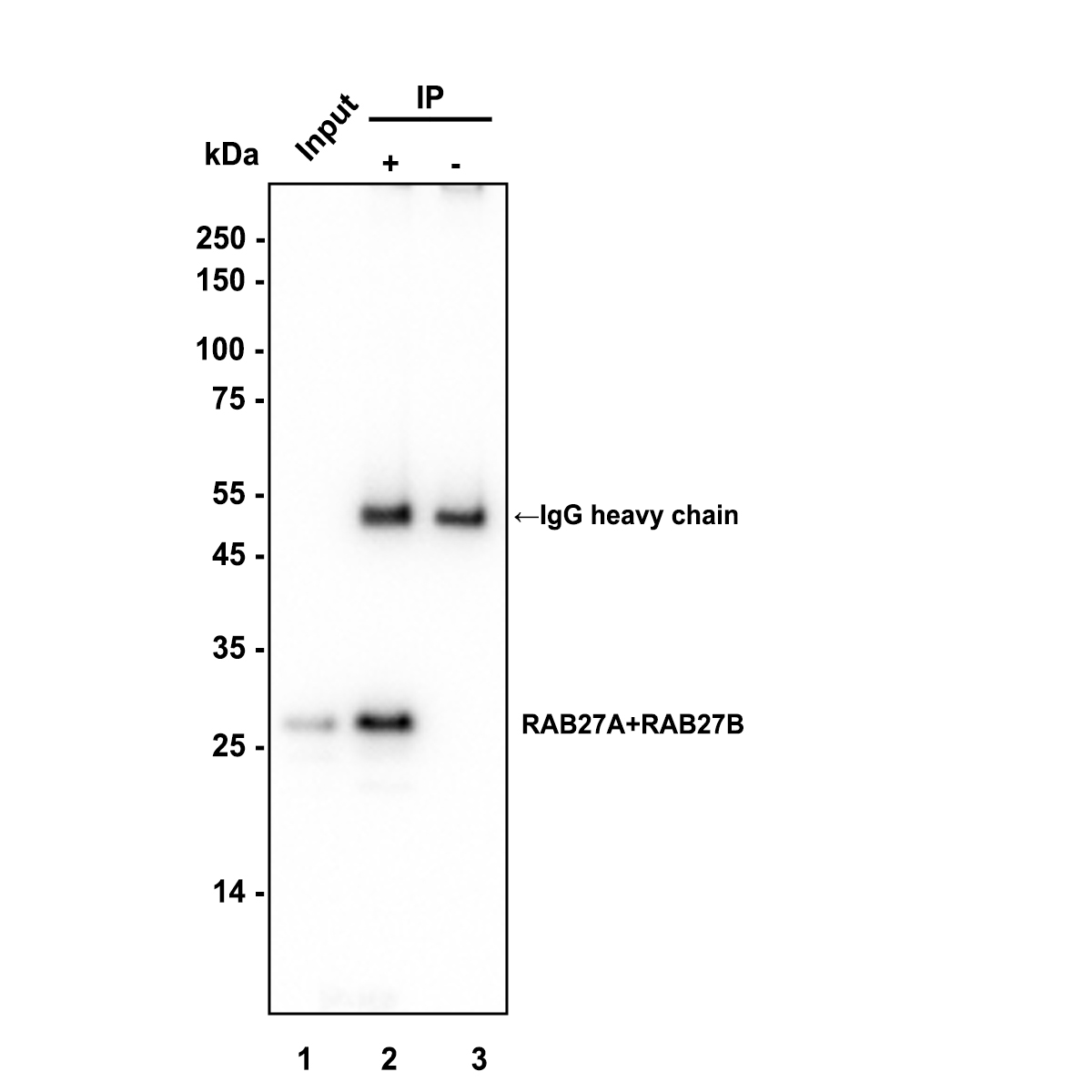
Immunoprecipitation (IP). RAB27A+RAB27B was immunoprecipitated in 0.2 mg K-562 (Human chronic myelogenous leukemia cell) cell lysate with RAB27A+RAB27B Recombinant Rabbit Monoclonal Antibodyat 2 µg/10 µl beads. Western blot was performed from the immunoprecipitate using RAB27A+RAB27B Recombinant Rabbit Monoclonal Antibodyat 1/5,000 dilution. Alpaca anti-Rabbit IgG Fc, Recombinant VHH monoclonal Antibody at 1/50000 dilution was used for 1 hour at room temperature. Lane 1: K-562 cell lysate (input). Lane 2: RAB27A+RAB27B Recombinant Rabbit Monoclonal Antibody IP in K-562 cell lysate. Lane 3: Rabbit IgG instead of RAB27A+RAB27B Recombinant Rabbit Monoclonal Antibody in K-562 cell lysate. Exposure time: 2 seconds. Blocking: 5% NFDM/TBST, 1 hour at room temperature. Primary dilution: RAB27A+RAB27B Recombinant Rabbit Monoclonal Antibody, 1/5,000 in primary antibody dilution buffer, 2 hours at room temperature.| Product Name | RAB27A+RAB27B Recombinant Rabbit Monoclonal Antibody |
|---|---|
| Antibody Type | Primary Antibodies |
| Immunogen | Recombinant protein within human RAB27A aa 1-221. |
| Clonality | Monoclonal |
|---|---|
| Isotype | IgG |
| Host Species | Rabbit |
| Tested Applications | FCIPWB |
| Species Reactivity | HumanMouseRat |
| Concentration | 1mg/ml |
| Purification | Protein A |
| Gene Symbol | RAB27A |
|---|---|
| Gene Synonyms | GS2 RAM RAB27 HsT18676 |
| Gene Full Name | RAB27A, member RAS oncogene family |
| Gene Summary | The protein encoded by this gene belongs to the small GTPase superfamily, Rab family. The protein is membrane-bound and may be involved in protein transport and small GTPase mediated signal transduction. Mutations in this gene are associated with Griscelli syndrome type 2. Alternative splicing occurs at this locus and four transcript variants encoding the same protein have been identified. [provided by RefSeq, Jul 2008] |
| Alternative Names | Ras-related protein Rab-27A EC:3.6.5.2 Rab-27 GTP-binding protein Ram RAB27A RAB27 Ras-related protein Rab-27B C25KG RAB27B |
| Molecular Weight(MW) | 25 kDa(Observed band size:27 kDa) |
| Cellular Localization | Membrane, Melanosome, Late endosome, Lysosome. |
WB
Western blot analysis of RAB27A+RAB27B on different lysates with Rabbit anti-RAB27A+RAB27B antibody at 1/5,000 dilution. Lysates/proteins at 15 µg/Lane. Exposure time: 10 seconds; Negative control: Raji. Blocking: 5% NFDM/TBST, 1 hour at room temperature. Primary antibody: RAB27A+RAB27B Recombinant Rabbit Monoclonal Antibody, 1/5,000 in primary antibody dilution buffer, overnight at 4 ℃. Secondary antibody: Goat anti-Rabbit IgG-HRP, 1/50,000 in 5% NFDM/TBST, 1 hour at room temperature.
FC
Application: Flow Cytometry (Intra). Species: Rat. Sample: PC-12 (Rat pheochromocytoma cells (undifferentiated)). Fixation: 4% Paraformaldehyde, 15 minutes at room temperature. Permeabilization: 1x Permeabilization Buffer (eBioscience), 15 minutes at room temperature. Blocking: 1% BSA + 10% normal goat serum, 15 minutes at room temperature. Antibody dilution buffer: 1x PBS. Primary antibody: RAB27A+RAB27B Recombinant Rabbit Monoclonal Antibody(1/1,000, Red) compared with Rabbit IgG Isotype Control (Green), 15 minutes at room temperature. Secondary antibody: Goat Anti-Rabbit IgG (488), 15 minutes at room temperature.
IP
Immunoprecipitation (IP). RAB27A+RAB27B was immunoprecipitated in 0.2 mg K-562 (Human chronic myelogenous leukemia cell) cell lysate with RAB27A+RAB27B Recombinant Rabbit Monoclonal Antibodyat 2 µg/10 µl beads. Western blot was performed from the immunoprecipitate using RAB27A+RAB27B Recombinant Rabbit Monoclonal Antibodyat 1/5,000 dilution. Alpaca anti-Rabbit IgG Fc, Recombinant VHH monoclonal Antibody at 1/50000 dilution was used for 1 hour at room temperature. Lane 1: K-562 cell lysate (input). Lane 2: RAB27A+RAB27B Recombinant Rabbit Monoclonal Antibody IP in K-562 cell lysate. Lane 3: Rabbit IgG instead of RAB27A+RAB27B Recombinant Rabbit Monoclonal Antibody in K-562 cell lysate. Exposure time: 2 seconds. Blocking: 5% NFDM/TBST, 1 hour at room temperature. Primary dilution: RAB27A+RAB27B Recombinant Rabbit Monoclonal Antibody, 1/5,000 in primary antibody dilution buffer, 2 hours at room temperature.| Form | Liquid |
|---|---|
| Storage Instructions | Shipped at 4℃. Store at +4℃ short term (1-2 weeks). Store at -20℃ long term. |
| Storage Buffer | 1*PBS (pH7.4), 0.1% BSA, 40% Glycerol, 0.2% Proclean 950. |


