WB
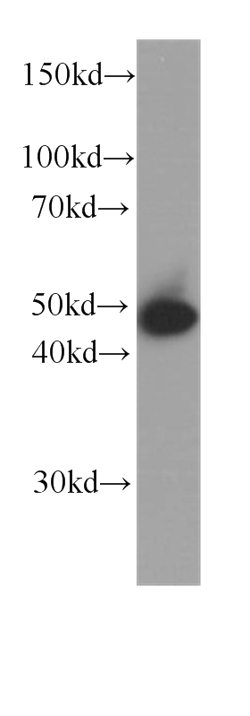
WB analysis of A431 using Cytokeratin 15 Monoclonal antibody. A431 cells were subjected to SDS PAGE followed by western blot with Cytokeratin 15 antibody at dilution of 1:1000 incubated at room temperature for 1.5 hours.IHC
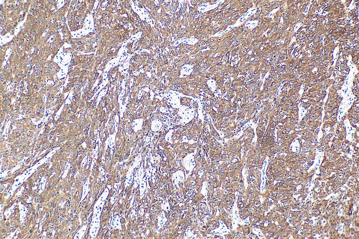
IHC staining of human cervical cancer using Cytokeratin 15 Monoclonal antibody. Immunohistochemical analysis of paraffin-embedded human cervical cancer tissue slide using Cytokeratin 15 antibody at dilution of 1:4000 (under 10x lens). Heat mediated antigen retrieval with Tris-EDTA buffer (pH 9.0).IHC
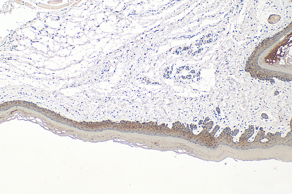
IHC staining of mouse skin using Cytokeratin 15 Monoclonal antibody. Immunohistochemical analysis of paraffin-embedded mouse skin tissue slide using Cytokeratin 15 antibody at dilution of 1:4000 (under 10x lens). Heat mediated antigen retrieval with Tris-EDTA buffer (pH 9.0).IHC
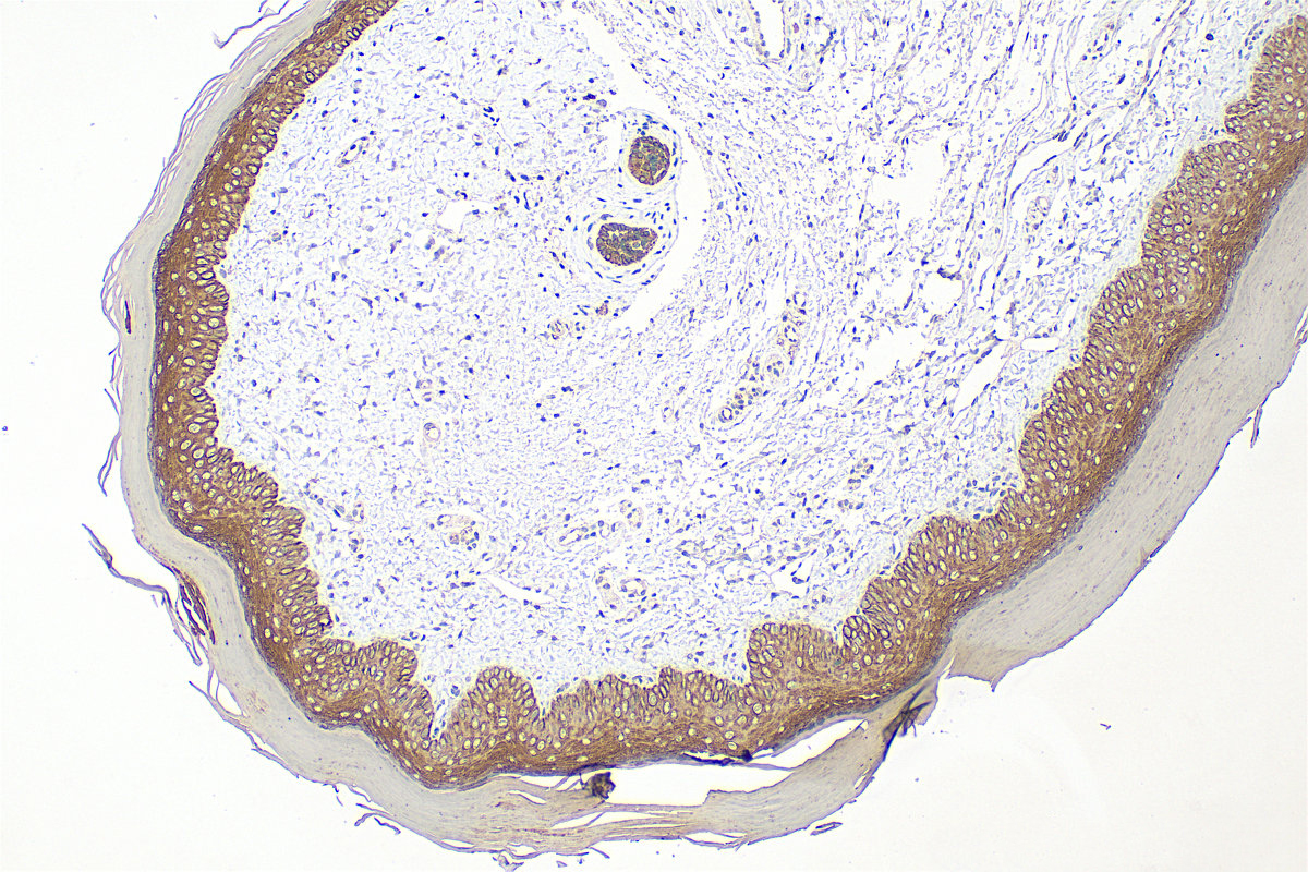
IHC staining of rat skin using Cytokeratin 15 Monoclonal antibody. Immunohistochemical analysis of paraffin-embedded rat skin tissue slide using Cytokeratin 15 antibody at dilution of 1:4000 (under 10x lens). Heat mediated antigen retrieval with Tris-EDTA buffer (pH 9.0).ICC/IF
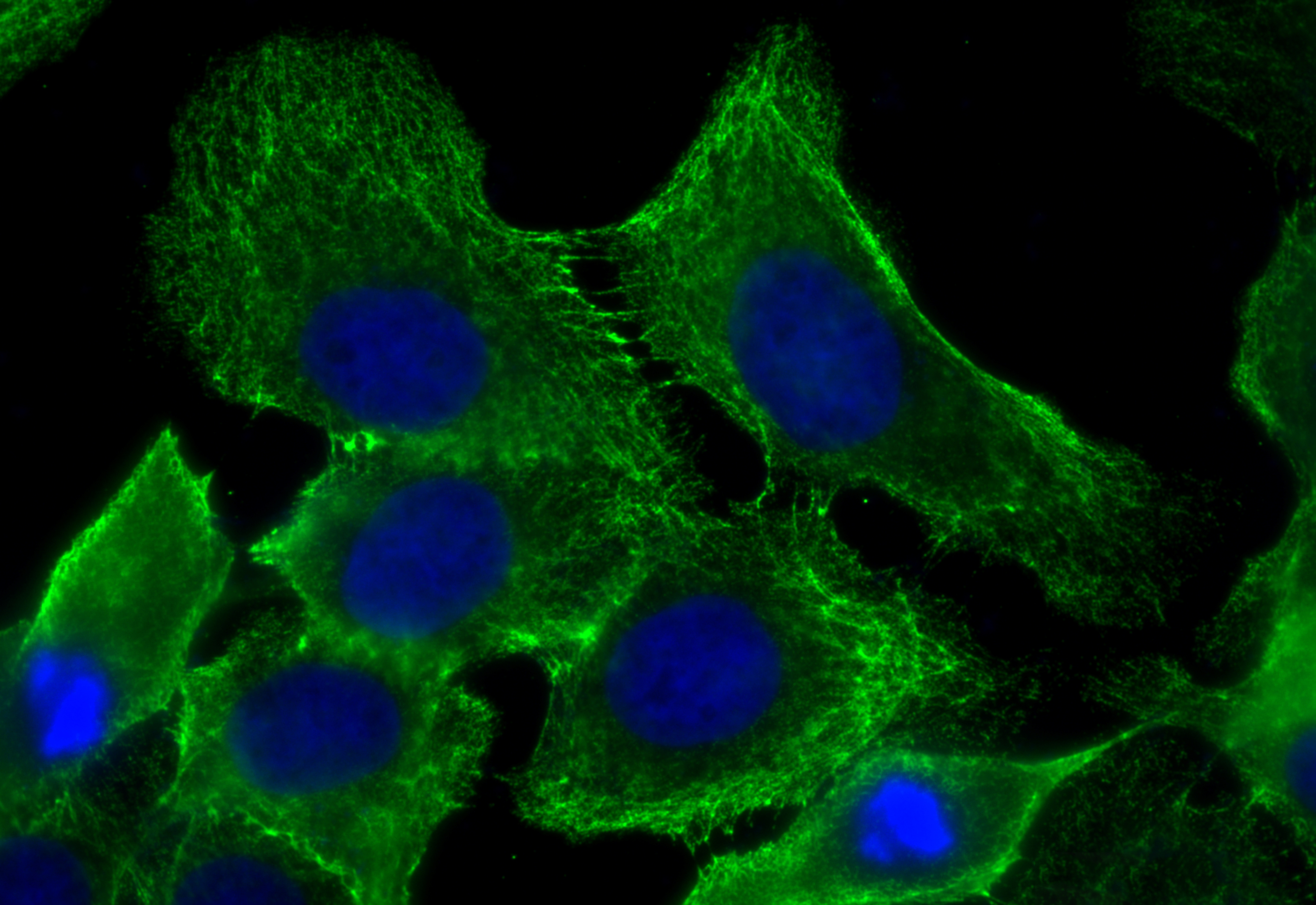
IF Staining of A431 using Cytokeratin 15 Monoclonal antibody. Immunofluorescent analysis of (-20°C Methanol) fixed A431 cells using Cytokeratin 15 antibody at dilution of 1:800 and 488-Conjugated Goat Anti-Mouse IgG(H+L).ICC/IF
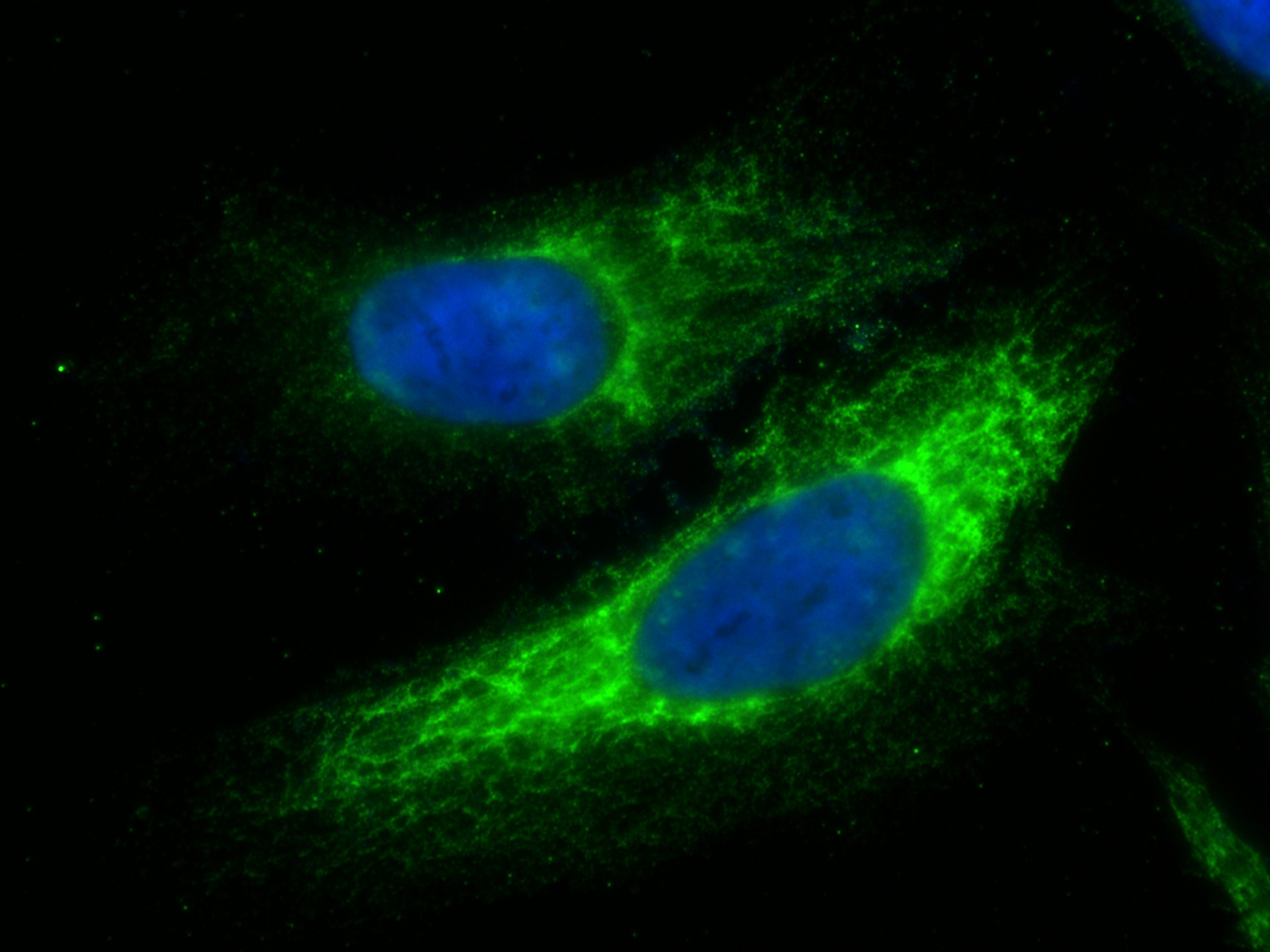
IF Staining of HeLa using Cytokeratin 15 Monoclonal antibody. Immunofluorescent analysis of (-20°C Methanol) fixed HeLa cells using Cytokeratin 15 antibody at dilution of 1:250 and 488-Conjugated Goat Anti-Mouse IgG(H+L).IF-P
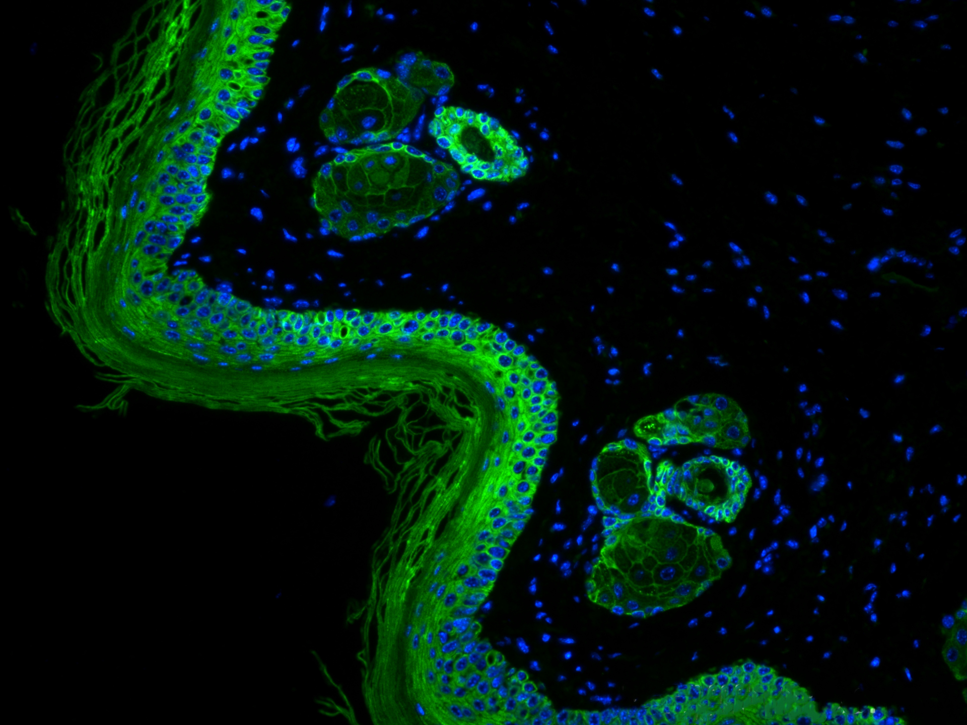
IF Staining of mouse skin using Cytokeratin 15 Monoclonal antibody. Immunofluorescent analysis of (4% PFA) fixed paraffin-embedded mouse skin tissue using Cytokeratin 15 antibody at dilution of 1:400 and 488-Conjugated Goat Anti-Mouse IgG(H+L) . Heat mediated antigen retrieval with Tris-EDTA buffer (pH 9.0).| Product Name | Cytokeratin 15 Monoclonal antibody |
|---|---|
| Antibody Type | Primary Antibodies |
| Immunogen | Recombinant human KRT15 protein. |
| Clonality | Monoclonal |
|---|---|
| Isotype | IgG1 |
| Host Species | Mouse |
| Tested Applications | ICC/IFIF-PIHCWB |
| WB:1:500-1:2000 IHC:1:2000-1:8000 ICC/IF:1:125-1:500 |
|
| Species Reactivity | HumanMouseRat |
| Concentration | 1mg/ml |
| Purification | Protein G |
| Gene Symbol | KRT15 |
|---|---|
| Gene Synonyms | K15 CK15 K1CO |
| Gene Full Name | keratin 15 |
| Gene Summary | The protein encoded by this gene is a member of the keratin gene family. The keratins are intermediate filament proteins responsible for the structural integrity of epithelial cells and are subdivided into cytokeratins and hair keratins. Most of the type I cytokeratins consist of acidic proteins which are arranged in pairs of heterotypic keratin chains and are clustered in a region on chromosome 17q21.2. [provided by RefSeq, Jul 2008] |
| Alternative Names | KRT15 6B4F8 CK 15 CK15 CK-15 |
| Molecular Weight(MW) | 49 kDa |
| Cellular Localization | Cytoplasm,Intermediate filament,Keratin. |
WB
WB analysis of A431 using Cytokeratin 15 Monoclonal antibody. A431 cells were subjected to SDS PAGE followed by western blot with Cytokeratin 15 antibody at dilution of 1:1000 incubated at room temperature for 1.5 hours.
IHC
IHC staining of human cervical cancer using Cytokeratin 15 Monoclonal antibody. Immunohistochemical analysis of paraffin-embedded human cervical cancer tissue slide using Cytokeratin 15 antibody at dilution of 1:4000 (under 10x lens). Heat mediated antigen retrieval with Tris-EDTA buffer (pH 9.0).
IHC
IHC staining of mouse skin using Cytokeratin 15 Monoclonal antibody. Immunohistochemical analysis of paraffin-embedded mouse skin tissue slide using Cytokeratin 15 antibody at dilution of 1:4000 (under 10x lens). Heat mediated antigen retrieval with Tris-EDTA buffer (pH 9.0).
IHC
IHC staining of rat skin using Cytokeratin 15 Monoclonal antibody. Immunohistochemical analysis of paraffin-embedded rat skin tissue slide using Cytokeratin 15 antibody at dilution of 1:4000 (under 10x lens). Heat mediated antigen retrieval with Tris-EDTA buffer (pH 9.0).
ICC/IF
IF Staining of A431 using Cytokeratin 15 Monoclonal antibody. Immunofluorescent analysis of (-20°C Methanol) fixed A431 cells using Cytokeratin 15 antibody at dilution of 1:800 and 488-Conjugated Goat Anti-Mouse IgG(H+L).
ICC/IF
IF Staining of HeLa using Cytokeratin 15 Monoclonal antibody. Immunofluorescent analysis of (-20°C Methanol) fixed HeLa cells using Cytokeratin 15 antibody at dilution of 1:250 and 488-Conjugated Goat Anti-Mouse IgG(H+L).
IF-P
IF Staining of mouse skin using Cytokeratin 15 Monoclonal antibody. Immunofluorescent analysis of (4% PFA) fixed paraffin-embedded mouse skin tissue using Cytokeratin 15 antibody at dilution of 1:400 and 488-Conjugated Goat Anti-Mouse IgG(H+L) . Heat mediated antigen retrieval with Tris-EDTA buffer (pH 9.0).| Application Notes | WB:1:500-1:2000 IHC:1:2000-1:8000 ICC/IF:1:125-1:500 |
|---|
| Form | Liquid |
|---|---|
| Storage Instructions | Store at -20°C. Stable for one year after shipment. Aliquoting is unnecessary for -20°C storage. |
| Storage Buffer | PBS with 0.02% sodium azide and 50% glycerol, pH 7.3. |


