WB
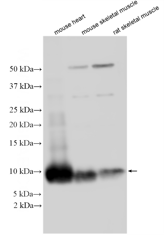
WB analysis using COX7C Polyclonal antibody. Various lysates were subjected to SDS PAGE followed by western blot with COX7C antibody at dilution of 1:1000 incubated at room temperature for 1.5 hours.IHC
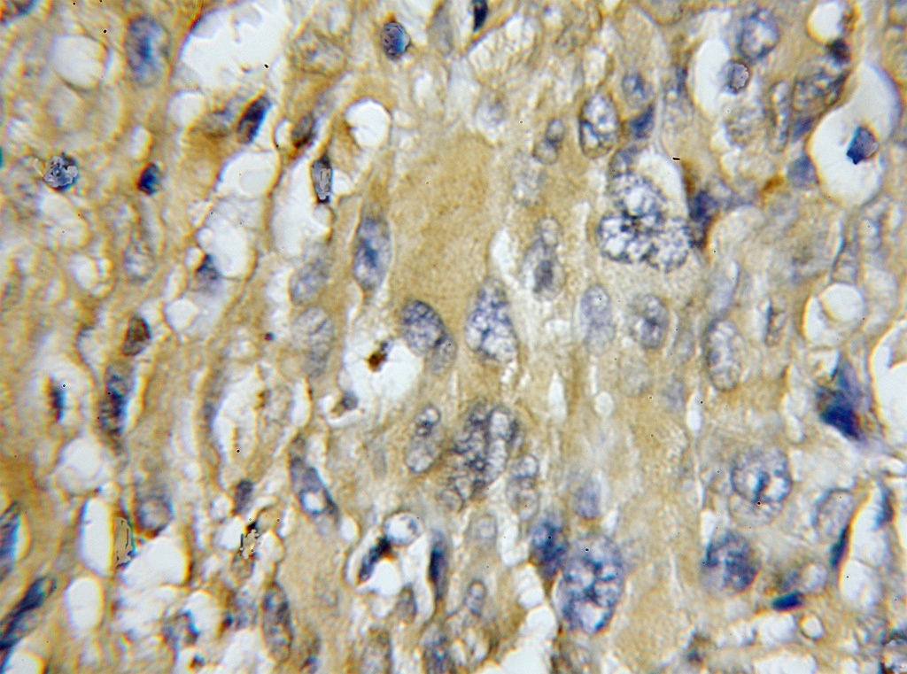
IHC staining of human gliomas using COX7C Polyclonal antibody. Immunohistochemical analysis of paraffin-embedded human gliomas using COX7C antibody at dilution of 1:100 (under 10x lens).IHC
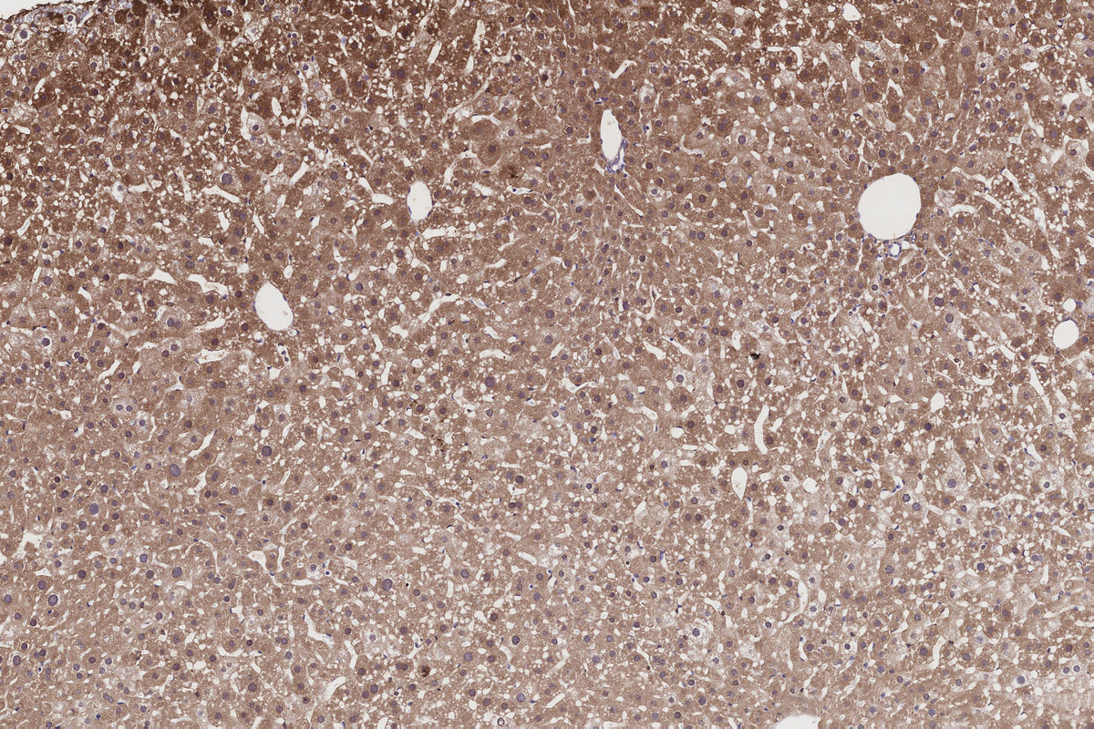
IHC staining of mouse liver using COX7C Polyclonal antibody. Immunohistochemical analysis of paraffin-embedded mouse liver tissue slide using COX7C antibody at dilution of 1:500 (under 10x lens). Heat mediated antigen retrieval with Tris-EDTA buffer (pH 9.0).IHC
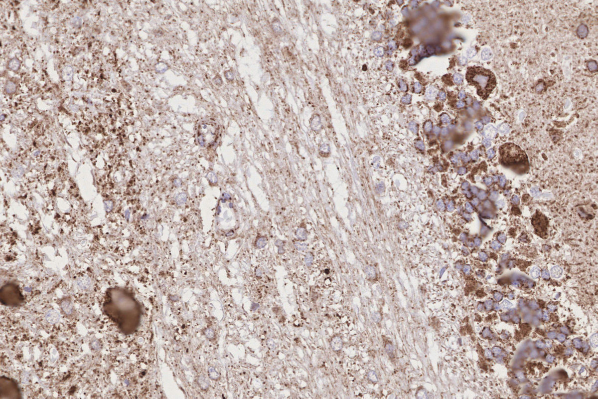
IHC staining of rat cerebellum using COX7C Polyclonal antibody. Immunohistochemical analysis of paraffin-embedded rat cerebellum tissue slide using COX7C antibody at dilution of 1:500 (under 40x lens). Heat mediated antigen retrieval with Tris-EDTA buffer (pH 9.0).ICC/IF
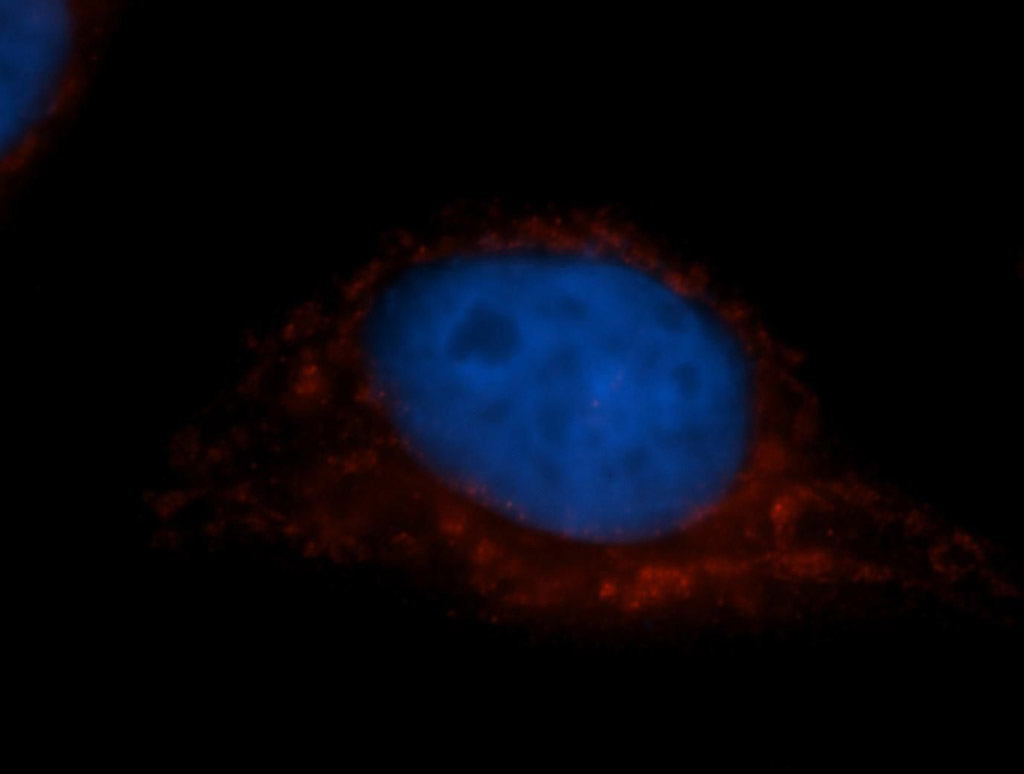
IF Staining of MCF-7 using COX7C Polyclonal antibody. Immunofluorescent analysis of MCF-7 cells, using COX7C antibody COX7C Polyclonal antibody at 1:50 dilution and Rhodamine-labeled goat anti-rabbit IgG (red). Blue pseudocolor = DAPI (fluorescent DNA dye).| Product Name | COX7C Polyclonal antibody |
|---|---|
| Antibody Type | Primary Antibodies |
| Immunogen | Recombinant human COX7C protein. |
| Clonality | Polyclonal |
|---|---|
| Isotype | IgG |
| Host Species | Rabbit |
| Tested Applications | ICC/IFIHCWB |
| WB:1:500-1:2000 IHC:1:20-1:200 ICC/IF:1:20-1:200 |
|
| Species Reactivity | HumanMouseRat |
| Concentration | 1mg/ml |
| Purification | Affinity purified |
| Gene Symbol | COX7C |
|---|---|
| Gene Full Name | cytochrome c oxidase subunit 7C |
| Gene Summary | Cytochrome c oxidase (COX), the terminal component of the mitochondrial respiratory chain, catalyzes the electron transfer from reduced cytochrome c to oxygen. This component is a heteromeric complex consisting of 3 catalytic subunits encoded by mitochondrial genes and multiple structural subunits encoded by nuclear genes. The mitochondrially-encoded subunits function in electron transfer, and the nuclear-encoded subunits may function in the regulation and assembly of the complex. This nuclear gene encodes subunit VIIc, which shares 87% and 85% amino acid sequence identity with mouse and bovine COX VIIc, respectively, and is found in all tissues. A pseudogene COX7CP1 has been found on chromosome 13. [provided by RefSeq, Jul 2008] |
| Alternative Names | Cytochrome c oxidase subunit 7C mitochondrial |
| Molecular Weight(MW) | 63 aa, 7 kDa(Observed band size: 15~28 kDa) |
| Cellular Localization | Membrane,Mitochondrion,Mitochondrion inner membrane. |
| SwissProt ID | P15954 |
|---|
WB
WB analysis using COX7C Polyclonal antibody. Various lysates were subjected to SDS PAGE followed by western blot with COX7C antibody at dilution of 1:1000 incubated at room temperature for 1.5 hours.
IHC
IHC staining of human gliomas using COX7C Polyclonal antibody. Immunohistochemical analysis of paraffin-embedded human gliomas using COX7C antibody at dilution of 1:100 (under 10x lens).
IHC
IHC staining of mouse liver using COX7C Polyclonal antibody. Immunohistochemical analysis of paraffin-embedded mouse liver tissue slide using COX7C antibody at dilution of 1:500 (under 10x lens). Heat mediated antigen retrieval with Tris-EDTA buffer (pH 9.0).
IHC
IHC staining of rat cerebellum using COX7C Polyclonal antibody. Immunohistochemical analysis of paraffin-embedded rat cerebellum tissue slide using COX7C antibody at dilution of 1:500 (under 40x lens). Heat mediated antigen retrieval with Tris-EDTA buffer (pH 9.0).
ICC/IF
IF Staining of MCF-7 using COX7C Polyclonal antibody. Immunofluorescent analysis of MCF-7 cells, using COX7C antibody COX7C Polyclonal antibody at 1:50 dilution and Rhodamine-labeled goat anti-rabbit IgG (red). Blue pseudocolor = DAPI (fluorescent DNA dye).| Application Notes | WB:1:500-1:2000 IHC:1:20-1:200 ICC/IF:1:20-1:200 |
|---|
| Form | Liquid |
|---|---|
| Storage Instructions | Store at -20°C. Stable for one year after shipment. Aliquoting is unnecessary for -20°C storage. |
| Storage Buffer | PBS with 0.02% sodium azide and 50% glycerol, pH 7.3. |


