WB
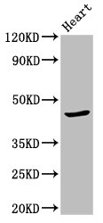
Western Blot. Positive WB detected in: Mouse heart tissue. All lanes: APOBEC3F antibody at 2.7μg/ml. Secondary Goat polyclonal to rabbit IgG at 1/50000 dilution.ICC/IF
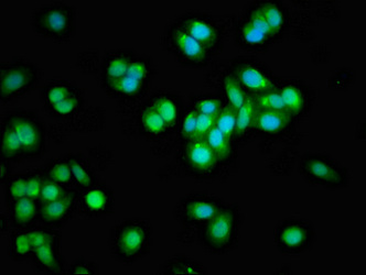
Immunofluorescent analysis of HepG2 cells using APOBEC3F Antibody at dilution of 1:100 and Alexa Fluor 488-congugated AffiniPure Goat Anti-Rabbit IgG(H+L).| Product Name | Rabbit anti-APOBEC3F Polyclonal antibody |
|---|---|
| Antibody Type | Primary Antibodies |
| Immunogen | Recombinant Human DNA dC->dU-editing enzyme APOBEC-3F protein (139-287AA). |
| Clonality | Polyclonal |
|---|---|
| Isotype | IgG |
| Host Species | Rabbit |
| Tested Applications | ICC/IFWB |
| WB:1:500-1:5000 ICC/IF:1:50-1:200 |
|
| Species Reactivity | HumanMouse |
| Concentration | 1mg/ml |
| Purification | Protein G |
| Gene Symbol | APOBEC3F |
|---|---|
| Gene Synonyms | A3F KA6 ARP8 BK150C2.4.MRNA |
| Gene Full Name | apolipoprotein B mRNA editing enzyme catalytic subunit 3F |
| Gene Summary | This gene is a member of the cytidine deaminase gene family. It is one of seven related genes or pseudogenes found in a cluster, thought to result from gene duplication, on chromosome 22. Members of the cluster encode proteins that are structurally and functionally related to the C to U RNA-editing cytidine deaminase APOBEC1. It is thought that the proteins may be RNA editing enzymes and have roles in growth or cell cycle control. Alternatively spliced transcript variants encoding different isoforms have been identified. [provided by RefSeq, Jul 2008] |
| Alternative Names | A3F antibody ABC3F_HUMAN antibody APOBEC3F antibody Apolipoprotein B mRNA editing enzyme catalytic polypeptide like 3F antibody Apolipoprotein B mRNA-editing enzyme catalytic polypeptide-like 3F antibody ARP8 antibody DNA dC->dU-editing enzyme APOBEC-3F antibody Induced upon T cell activation antibody KA6 antibody |
| Molecular Weight(MW) | 46, 10, 12 kDa(Observed band size: 46 kDa) |
| Cellular Localization | Cytoplasm. |
| SwissProt ID | Q8IUX4 |
|---|
WB
Western Blot. Positive WB detected in: Mouse heart tissue. All lanes: APOBEC3F antibody at 2.7μg/ml. Secondary Goat polyclonal to rabbit IgG at 1/50000 dilution.
ICC/IF
Immunofluorescent analysis of HepG2 cells using APOBEC3F Antibody at dilution of 1:100 and Alexa Fluor 488-congugated AffiniPure Goat Anti-Rabbit IgG(H+L).| Application Notes | WB:1:500-1:5000 ICC/IF:1:50-1:200 |
|---|
| Form | Liquid |
|---|---|
| Storage Instructions | Upon receipt, store at -20°C or -80°C. Avoid repeated freeze. |
| Storage Buffer | Preservative: 0.03% Proclin 300; Constituents: 50% Glycerol, 0.01M PBS, pH 7.4. |


