WB
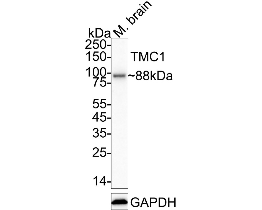
Western blot analysis of TMC1 on mouse brain tissue lysates with Rabbit anti-TMC1 antibody at 1/2,000 dilution. Lysates/proteins at 40 µg/Lane. Exposure time: 3 minutes; 4-20% SDS-PAGE gel. Proteins were transferred to a PVDF membrane and blocked with 5% NFDM/TBST for 1 hour at room temperature. The primary antibody at 1/2,000 dilution was used in 5% NFDM/TBST at 4℃ overnight. Goat Anti-Rabbit IgG - HRP Secondary Antibody at 1/50,000 dilution was used for 1 hour at room temperature.WB
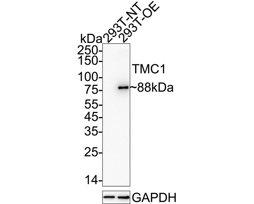
Western blot analysis of TMC1 on different lysates with Rabbit anti-TMC1 antibody at 1/2,000 dilution. Lysates/proteins at 15 µg/Lane. Exposure time: 3 minutes; 4-20% SDS-PAGE gel. Proteins were transferred to a PVDF membrane and blocked with 5% NFDM/TBST for 1 hour at room temperature. The primary antibody at 1/2,000 dilution was used in 5% NFDM/TBST at 4℃ overnight. Goat Anti-Rabbit IgG - HRP Secondary Antibody at 1/50,000 dilution was used for 1 hour at room temperature.ICC/IF
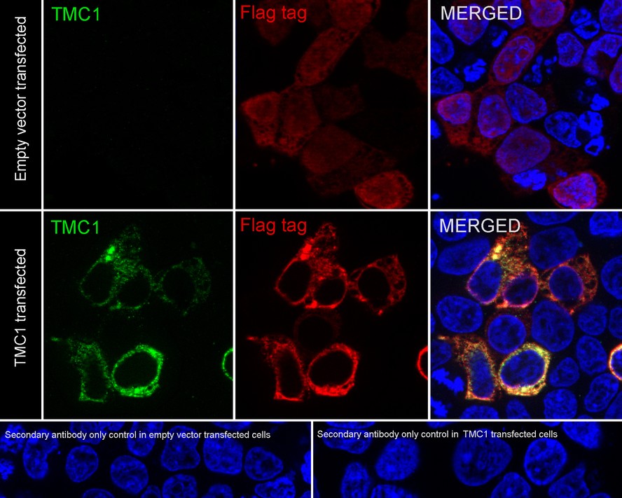
Immunocytochemistry analysis of 293T cells labeling TMC1 with Rabbit anti-TMC1 antibody at 1/2,000 dilution. 293T cells, transfected with empty control (top, negative) / Flag-tagged TMC1 (bottom, positive) expression vector, respectively, were fixed in 4% paraformaldehyde for 15 minutes at room temperature, permeabilized with 0.1% Triton X-100 in PBS for 15 minutes at room temperature, then blocked with 1% BSA in 10% negative goat serum for 1 hour at room temperature. Cells were then incubated with Rabbit anti-TMC1 antibody at 1/2,000 dilution in 1% BSA in PBST overnight at 4 ℃. Goat Anti-Rabbit IgG H&L (488) was used as the secondary antibody at 1/1,000 dilution. PBS instead of the primary antibody was used as the secondary antibody only control. Nuclear DNA was labelled in blue with DAPI. Flag (red) was stained at 1/100 dilution overnight at +4℃. Goat Anti-Mouse IgG H&L (594) was used as the secondary antibody at 1/1,000 dilution.| Product Name | TMC1 Recombinant Rabbit Monoclonal Antibody |
|---|---|
| Antibody Type | Primary Antibodies |
| Immunogen | Recombinant protein within human TMC1 aa 1-150. |
| Clonality | Monoclonal |
|---|---|
| Isotype | IgG |
| Host Species | Rabbit |
| Tested Applications | ICC/IFWB |
| WB:1:2000 ICC/IF:1:2000 |
|
| Species Reactivity | HumanMouse |
| Concentration | 1mg/ml |
| Purification | Protein A |
| Gene Symbol | TMC1 |
|---|---|
| Gene Synonyms | DFNB7 DFNA36 DFNB11 |
| Gene Full Name | transmembrane channel like 1 |
| Gene Summary | This gene is considered a member of a gene family predicted to encode transmembrane proteins. The specific function of this gene is unknown; however, it is known to be required for normal function of cochlear hair cells. Mutations in this gene have been associated with progressive postlingual hearing loss and profound prelingual deafness. [provided by RefSeq, Jul 2008] |
| Alternative Names | Transmembrane channel-like protein 1 1 publication Alternative names Transmembrane cochlear-expressed protein 1 Gene names Name TMC1 |
| Molecular Weight(MW) | 88 kDa |
| Cellular Localization | Cell membrane. |
| SwissProt ID | Q8TDI8 |
|---|
WB
Western blot analysis of TMC1 on mouse brain tissue lysates with Rabbit anti-TMC1 antibody at 1/2,000 dilution. Lysates/proteins at 40 µg/Lane. Exposure time: 3 minutes; 4-20% SDS-PAGE gel. Proteins were transferred to a PVDF membrane and blocked with 5% NFDM/TBST for 1 hour at room temperature. The primary antibody at 1/2,000 dilution was used in 5% NFDM/TBST at 4℃ overnight. Goat Anti-Rabbit IgG - HRP Secondary Antibody at 1/50,000 dilution was used for 1 hour at room temperature.
WB
Western blot analysis of TMC1 on different lysates with Rabbit anti-TMC1 antibody at 1/2,000 dilution. Lysates/proteins at 15 µg/Lane. Exposure time: 3 minutes; 4-20% SDS-PAGE gel. Proteins were transferred to a PVDF membrane and blocked with 5% NFDM/TBST for 1 hour at room temperature. The primary antibody at 1/2,000 dilution was used in 5% NFDM/TBST at 4℃ overnight. Goat Anti-Rabbit IgG - HRP Secondary Antibody at 1/50,000 dilution was used for 1 hour at room temperature.
ICC/IF
Immunocytochemistry analysis of 293T cells labeling TMC1 with Rabbit anti-TMC1 antibody at 1/2,000 dilution. 293T cells, transfected with empty control (top, negative) / Flag-tagged TMC1 (bottom, positive) expression vector, respectively, were fixed in 4% paraformaldehyde for 15 minutes at room temperature, permeabilized with 0.1% Triton X-100 in PBS for 15 minutes at room temperature, then blocked with 1% BSA in 10% negative goat serum for 1 hour at room temperature. Cells were then incubated with Rabbit anti-TMC1 antibody at 1/2,000 dilution in 1% BSA in PBST overnight at 4 ℃. Goat Anti-Rabbit IgG H&L (488) was used as the secondary antibody at 1/1,000 dilution. PBS instead of the primary antibody was used as the secondary antibody only control. Nuclear DNA was labelled in blue with DAPI. Flag (red) was stained at 1/100 dilution overnight at +4℃. Goat Anti-Mouse IgG H&L (594) was used as the secondary antibody at 1/1,000 dilution.| Application Notes | WB:1:2000 ICC/IF:1:2000 |
|---|
| Form | Liquid |
|---|---|
| Storage Instructions | Shipped at 4℃. Store at +4℃ short term (1-2 weeks). Store at -20℃ long term. |
| Storage Buffer | 1*PBS (pH7.4), 0.1% BSA, 40% Glycerol, 0.2% Proclean 950. |


