WB
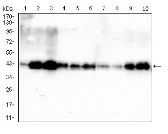
Western blot analysis using PDHA1 mouse mAb against HepG2 (1), HEK293 (2), HL-60 (3), SK-OV-3 (4), PC-3 (5), PANC-1 (6), NRK (7), C2C12 (8), C6 (9) and PC-12 (10) cell lysate.IHC
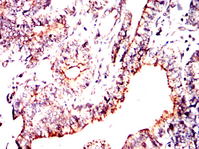
Immunohistochemical analysis of paraffin-embedded human lung cancer tissues using PDHA1 mouse mAb with DAB staining.IHC
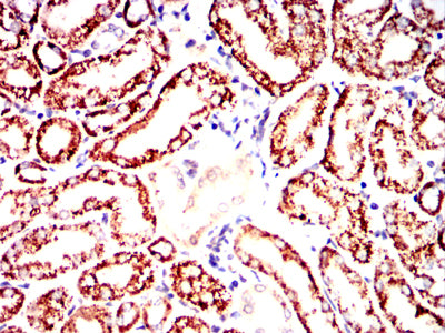
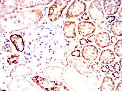
Immunohistochemical analysis of paraffin-embedded mouse kidney tissues using PDHA1 mouse mAb with DAB staining.IHC
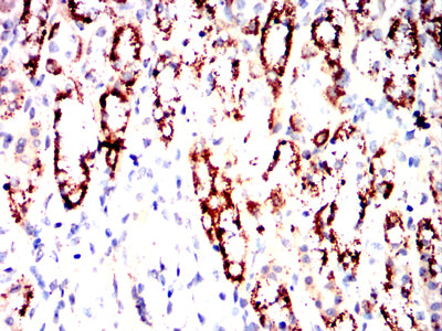
Immunohistochemical analysis of paraffin-embedded Rat kidney tissues using PDHA1 mouse mAb with DAB staining.IHC
Immunohistochemical analysis of paraffin-embedded Rabbit kidney tissues using PDHA1 mouse mAb with DAB staining.ICC/IF
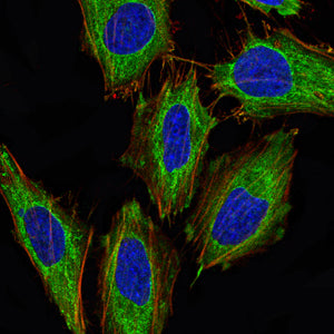
Immunofluorescence analysis of Hela cells using PDHA1 mouse mAb (green). Blue: DRAQ5 fluorescent DNA dye. Red: Actin filaments have been labeled with Alexa Fluor- 555 phalloidin. Secondary antibody from Fisher.FC
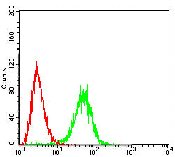
Flow cytometric analysis of Hela cells using PDHA1 mouse mAb (green) and negative control (red).| Product Name | Mouse Monoclonal Antibody to PDHA1 |
|---|---|
| Antibody Type | Primary Antibodies |
| Immunogen | Purified recombinant fragment of human PDHA1(AA: 241-390) expressed in E. Coli. |
| Clonality | Monoclonal |
|---|---|
| Isotype | IgG1 |
| Host Species | Mouse |
| Tested Applications | FCICC/IFIHCIPWB |
| WB:1:500 - 1:2000 IHC:1:200 - 1:1000 ICC/IF:1:50 - 1:200 FC:1:200 - 1:400 IP:1:200-1:300 |
|
| Species Reactivity | HumanMouseRat |
| Concentration | 1mg/ml |
| Purification | Affinity purified |
| Gene Symbol | PDHA1 |
|---|---|
| Gene Synonyms | PDHA PDHAD PHE1A E1alpha PDHCE1A |
| Gene Full Name | pyruvate dehydrogenase E1 subunit alpha 1 |
| Gene Summary | The pyruvate dehydrogenase (PDH) complex is a nuclear-encoded mitochondrial multienzyme complex that catalyzes the overall conversion of pyruvate to acetyl-CoA and CO(2), and provides the primary link between glycolysis and the tricarboxylic acid (TCA) cycle. The PDH complex is composed of multiple copies of three enzymatic components: pyruvate dehydrogenase (E1), dihydrolipoamide acetyltransferase (E2) and lipoamide dehydrogenase (E3). The E1 enzyme is a heterotetramer of two alpha and two beta subunits. This gene encodes the E1 alpha 1 subunit containing the E1 active site, and plays a key role in the function of the PDH complex. Mutations in this gene are associated with pyruvate dehydrogenase E1-alpha deficiency and X-linked Leigh syndrome. Alternatively spliced transcript variants encoding different isoforms have been found for this gene.[provided by RefSeq, Mar 2010] |
| Alternative Names | PDHA PDHAD PHE1A PDHCE1A |
| Molecular Weight(MW) | 43 kDa |
| Cellular Localization | Mitochondrion. |
| SwissProt ID | P08559 |
|---|
WB
Western blot analysis using PDHA1 mouse mAb against HepG2 (1), HEK293 (2), HL-60 (3), SK-OV-3 (4), PC-3 (5), PANC-1 (6), NRK (7), C2C12 (8), C6 (9) and PC-12 (10) cell lysate.
IHC
Immunohistochemical analysis of paraffin-embedded human lung cancer tissues using PDHA1 mouse mAb with DAB staining.
IHC
Immunohistochemical analysis of paraffin-embedded mouse kidney tissues using PDHA1 mouse mAb with DAB staining.
IHC
Immunohistochemical analysis of paraffin-embedded Rat kidney tissues using PDHA1 mouse mAb with DAB staining.
IHC
Immunohistochemical analysis of paraffin-embedded Rabbit kidney tissues using PDHA1 mouse mAb with DAB staining.
ICC/IF
Immunofluorescence analysis of Hela cells using PDHA1 mouse mAb (green). Blue: DRAQ5 fluorescent DNA dye. Red: Actin filaments have been labeled with Alexa Fluor- 555 phalloidin. Secondary antibody from Fisher.
FC
Flow cytometric analysis of Hela cells using PDHA1 mouse mAb (green) and negative control (red).| Application Notes | WB:1:500 - 1:2000 IHC:1:200 - 1:1000 ICC/IF:1:50 - 1:200 FC:1:200 - 1:400 IP:1:200-1:300 |
|---|
| Form | Liquid |
|---|---|
| Storage Instructions | Store at 4°C short term. Aliquot and store at -20°C long term. Avoid freeze/thaw cycles. |
| Storage Buffer | Purified antibody in PBS with 0.05% sodium azide. |


