WB
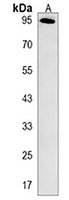
Western blot analysis of ARMC5 expression in mouse kidney (A) whole cell lysates.IHC
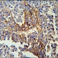
Immunohistochemical analysis of ARMC5 staining in human skin carcinoma formalin fixed paraffin embedded tissue section. The section was pre-treated using heat mediated antigen retrieval with sodium citrate buffer (pH 6.0). The section was then incubated with the antibody at room temperature and detected using an HRP conjugated compact polymer system. DAB was used as the chromogen. The section was then counterstained with haematoxylin and mounted with DPX.FC
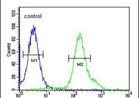
Flow cytometric analysis of 293 cells using Anti-ARMC5 Antibody. The cells were fixed with 2% paraformaldehyde (10 min) and then permeabilized with 90% methanol for 10 min. The cells were incubated in 2% bovine serum albumin to block non-specific protein-protein interactions followed by the antibody at 37 °C for 60 min. The secondary antibody Goat Anti-Rabbit IgG (H&L) - AF488 was incubated at 37 °C for 40 min. Isotype control antibody (blue line) was used under the same condition.| Product Name | Anti-ARMC5 Antibody |
|---|---|
| Antibody Type | Primary Antibodies |
| Immunogen | KLH-conjugated synthetic peptide encompassing a sequence within the C-terminal region of human ARMC5. The exact sequence is proprietary. |
| Clonality | Polyclonal |
|---|---|
| Isotype | IgG |
| Host Species | Rabbit |
| Tested Applications | FCIHCWB |
| WB:1:500-1:1000 IHC:1:50-1:200 FC:1:10-1:50 |
|
| Species Reactivity | HumanMouse |
| Concentration | 1mg/ml |
| Purification | Affinity purified |
| Gene Symbol | ARMC5 |
|---|---|
| Gene Synonyms | AIMAH2 |
| Gene Full Name | armadillo repeat containing 5 |
| Gene Summary | This gene encodes a member of the ARM (armadillo/beta-catenin-like repeat) superfamily. The ARM repeat is a tandemly repeated sequence motif with approximately 40 amino acid long. This repeat is implicated in mediating protein-protein interactions. The encoded protein contains seven ARM repeats. Mutations in this gene are associated with primary bilateral macronodular adrenal hyperplasia, which is also known as ACTH-independent macronodular adrenal hyperplasia 2. Alternatively spliced transcript variants encoding different isoforms have been found for this gene. [provided by RefSeq, Sep 2014] |
| Alternative Names | Armadillo repeat-containing protein 5 |
| Molecular Weight(MW) | 97 kDa(Observed band size: 95 kDa) |
| Cellular Localization | Chromosome,Cytoplasm,Nucleus. |
| SwissProt ID | Q96C12 |
|---|
WB
Western blot analysis of ARMC5 expression in mouse kidney (A) whole cell lysates.
IHC
Immunohistochemical analysis of ARMC5 staining in human skin carcinoma formalin fixed paraffin embedded tissue section. The section was pre-treated using heat mediated antigen retrieval with sodium citrate buffer (pH 6.0). The section was then incubated with the antibody at room temperature and detected using an HRP conjugated compact polymer system. DAB was used as the chromogen. The section was then counterstained with haematoxylin and mounted with DPX.
FC
Flow cytometric analysis of 293 cells using Anti-ARMC5 Antibody. The cells were fixed with 2% paraformaldehyde (10 min) and then permeabilized with 90% methanol for 10 min. The cells were incubated in 2% bovine serum albumin to block non-specific protein-protein interactions followed by the antibody at 37 °C for 60 min. The secondary antibody Goat Anti-Rabbit IgG (H&L) - AF488 was incubated at 37 °C for 40 min. Isotype control antibody (blue line) was used under the same condition.| Application Notes | WB:1:500-1:1000 IHC:1:50-1:200 FC:1:10-1:50 |
|---|
| Form | Liquid |
|---|---|
| Storage Instructions | Shipped at 4°C. Upon delivery aliquot and store at -20°C for one year. Avoid freeze/thaw cycles. |
| Storage Buffer | Liquid in 0.42% Potassium phosphate, 0.87% Sodium chloride, pH 7.3, 30% glycerol, and 0.01% sodium azide. |


