WB
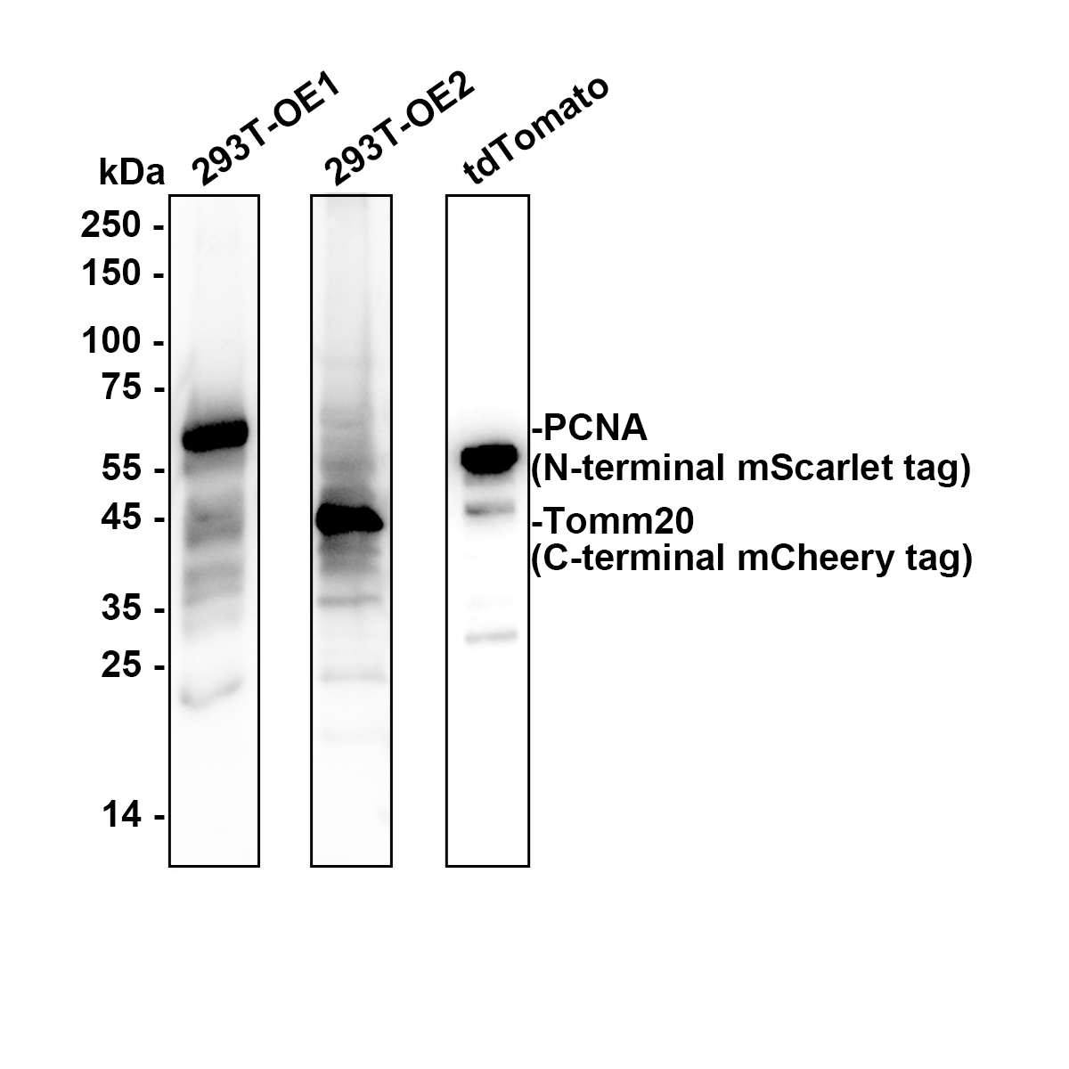
Western blot analysis of pan-RFP on different lysates with Rabbit anti-pan-RFP antibody at 1/10,000 dilution. Exposure time: 2 seconds; 4-20% SDS-PAGE gel. Proteins were transferred to a PVDF membrane and blocked with 5% NFDM/TBST for 1 hour at room temperature. The primary antibody at 1/10,000 dilution was used in K1803 at room temperature. Goat Anti-Rabbit IgG - HRP Secondary Antibody at 1/50,000 dilution was used for 1 hour at room temperature.ICC/IF
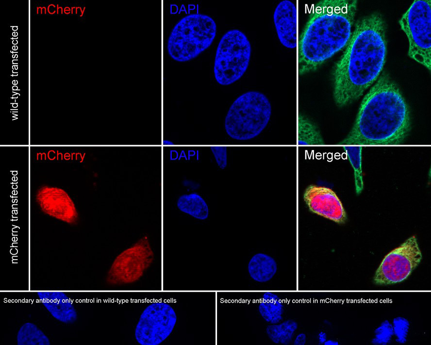
Application: Immunocytochemistry (IF-cell). Species: Human. Sample: HeLa (Human cervix adenocarcinoma epithelial cell) transfected with or without mCherry. Fixation: 4% Paraformaldehyde, 15 minutes at room temperature. Permeabilization: 0.1% Triton X-100, 15 minutes at room temperature. Blocking: 1% BSA + 10% normal goat serum, 1 hour at room temperature. Antibody dilution buffer: 1% BSA in PBST. Primary antibody: pan-RFP Rabbit Polyclonal Antibody, 1/2,000, overnight at 4℃. Secondary antibody: Goat Anti-Rabbit IgG (488), 45 minutes at room temperature. Counterstain: Beta tubulin (Red), 1/100, overnight at 4℃. The nuclear counterstain was DAPI (Blue).| Product Name | pan-RFP Rabbit Polyclonal Antibody |
|---|---|
| Antibody Type | Primary Antibodies |
| Immunogen | Recombinant protein within mCherry 1-236aa. |
| Clonality | Polyclonal |
|---|---|
| Isotype | IgG |
| Host Species | Rabbit |
| Tested Applications | ICC/IFWB |
| WB:1:10000-1:50000 ICC/IF:1:2000-1:5000 |
|
| Species Reactivity | All |
| Concentration | 1mg/ml |
| Purification | Affinity purified |
| Function | Red fluorescent proteins (RFPs) is a collective term referring to a heterogenous group of red chromophore-carrying proteins, originating from various species and forming different protein lineages. The original RFP (dsRed) is a 225 amino acid fluorescent protein (25.9 kDa) derived from Discosoma sp.. It emits red light with a peak wavelength of 593 nm upon excitation by green light (excitation peak at 558 nm). When fused with other proteins, RFP serves as a versatile reporter protein e.g. for quantifying expression levels or facilitates visualization of subcellular localization through fluorescence microscopy. |
|---|
WB
Western blot analysis of pan-RFP on different lysates with Rabbit anti-pan-RFP antibody at 1/10,000 dilution. Exposure time: 2 seconds; 4-20% SDS-PAGE gel. Proteins were transferred to a PVDF membrane and blocked with 5% NFDM/TBST for 1 hour at room temperature. The primary antibody at 1/10,000 dilution was used in K1803 at room temperature. Goat Anti-Rabbit IgG - HRP Secondary Antibody at 1/50,000 dilution was used for 1 hour at room temperature.
ICC/IF
Application: Immunocytochemistry (IF-cell). Species: Human. Sample: HeLa (Human cervix adenocarcinoma epithelial cell) transfected with or without mCherry. Fixation: 4% Paraformaldehyde, 15 minutes at room temperature. Permeabilization: 0.1% Triton X-100, 15 minutes at room temperature. Blocking: 1% BSA + 10% normal goat serum, 1 hour at room temperature. Antibody dilution buffer: 1% BSA in PBST. Primary antibody: pan-RFP Rabbit Polyclonal Antibody, 1/2,000, overnight at 4℃. Secondary antibody: Goat Anti-Rabbit IgG (488), 45 minutes at room temperature. Counterstain: Beta tubulin (Red), 1/100, overnight at 4℃. The nuclear counterstain was DAPI (Blue).| Application Notes | WB:1:10000-1:50000 ICC/IF:1:2000-1:5000 |
|---|
| Form | Liquid |
|---|---|
| Storage Instructions | Shipped at 4℃. Store at +4℃ short term (1-2 weeks). Store at -20℃ long term. |
| Storage Buffer | 1*PBS (pH7.4), 0.1% BSA, 40% Glycerol, 0.2% Proclean 950. |


