WB
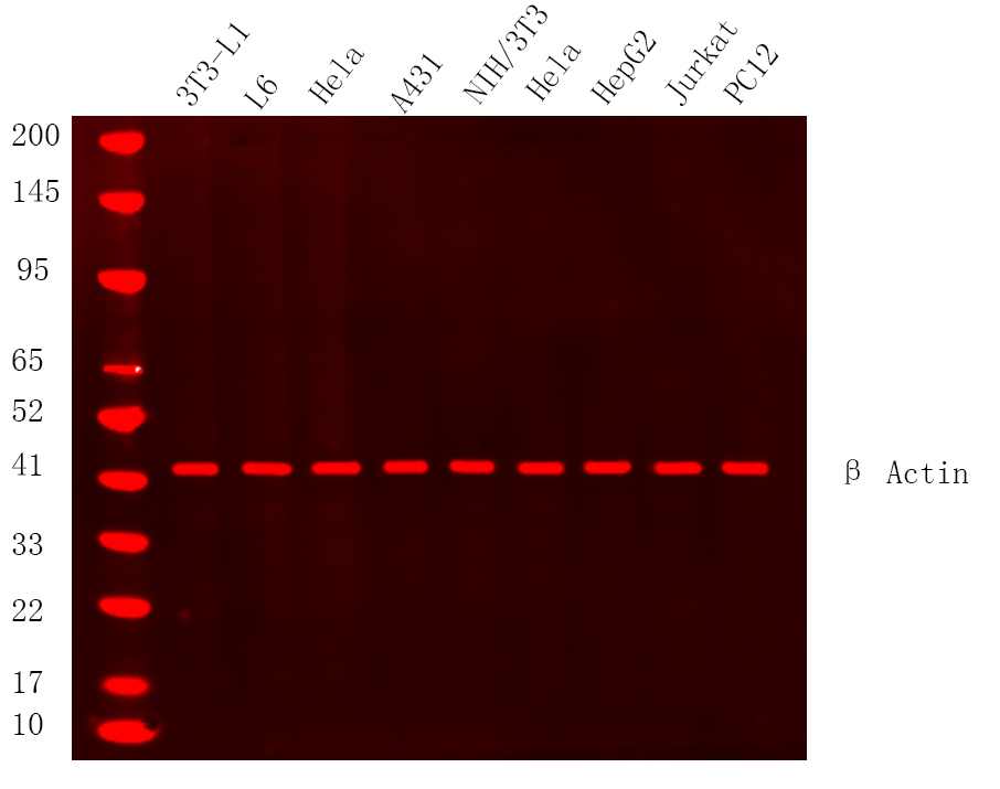
Western Blot analysis using Various whole cell lysates were separated by 4-20% SDS-PAGE, and the membrane was blotted with antiβ Actin Rabbit mAb diluted at 1:20000.WB
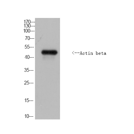
Western Blot analysis of Drosophilidae cells using Actin beta rabbit pAb (Drosophilidae). Secondary antibody was diluted at 1:20000.IHC
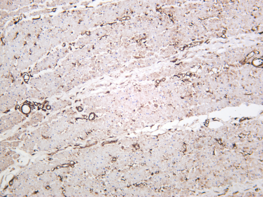
Human colon was stained with anti-β Actin rabbit antibody.IHC
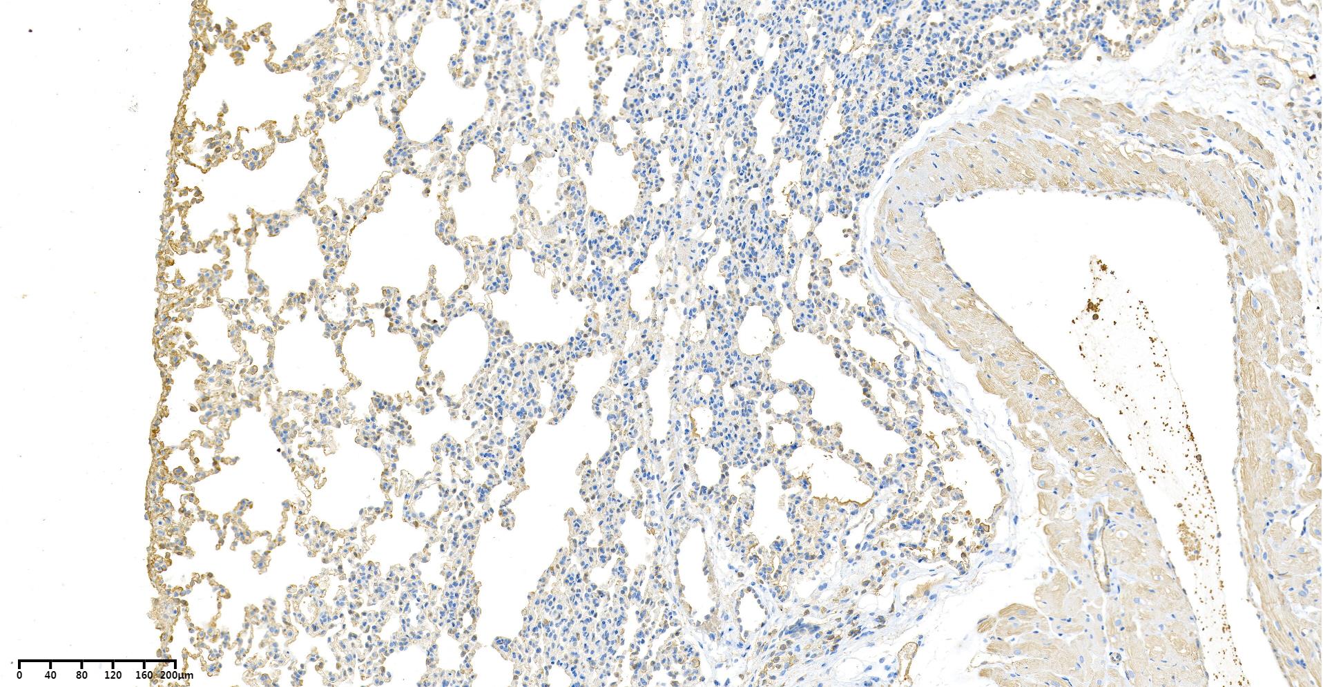
Rat lungs was stained with anti-β Actin Rabbit antibody.ICC/IF
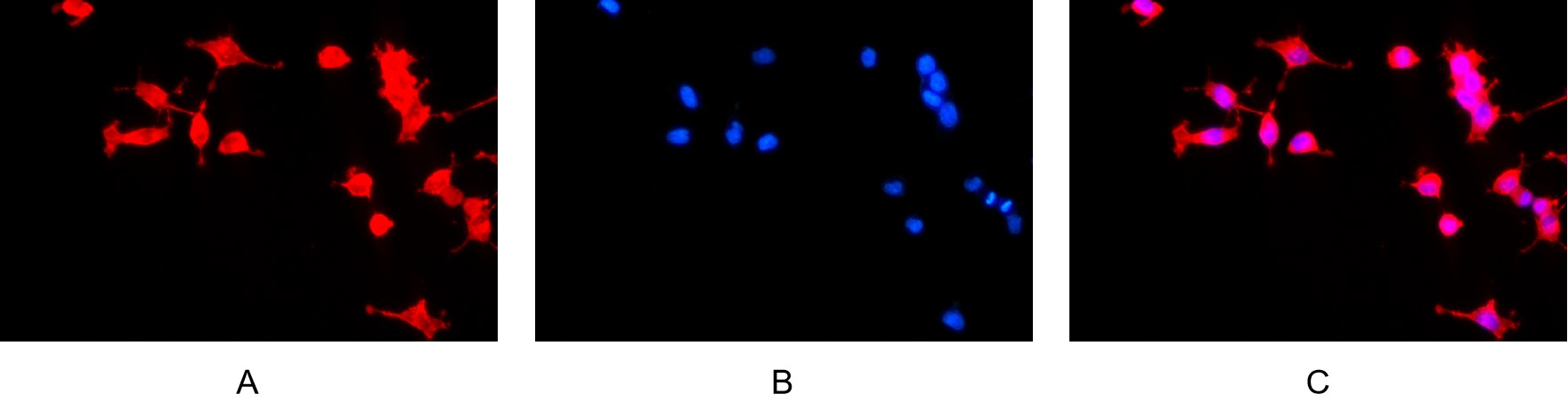
Immunofluorescence analysis of HEK293. Picture A: β Actin antibody (red). Picture B: DAPI (blue). Picture C: Merge of A+B.| Product Name | β-actin Rabbit mAb |
|---|---|
| Antibody Type | Primary Antibodies |
| Immunogen | The specific immunogen used to produce this antibody is proprietary information. |
| Clonality | Monoclonal |
|---|---|
| Isotype | IgG,K |
| Host Species | Rabbit |
| Tested Applications | ICC/IFIHCWB |
| WB:1:2000-1:10000 IHC:1:2000-1:20000 ICC/IF:1:200-1:1000 |
|
| Species Reactivity | BirdBovineCarpCatChickenDogFishHamsterHumanMousePigRainbow TroutRatSheep |
| Concentration | 1mg/ml |
| Purification | Protein A |
| Gene Symbol | ACTB |
|---|---|
| Gene Synonyms | BNS CSMH DDS1 THC8 BKRNS BRWS1 PS1TP5BP1 |
| Gene Full Name | actin beta |
| Gene Summary | This gene encodes one of six different actin proteins. Actins are highly conserved proteins that are involved in cell motility, structure, integrity, and intercellular signaling. The encoded protein is a major constituent of the contractile apparatus and one of the two nonmuscle cytoskeletal actins that are ubiquitously expressed. Mutations in this gene cause Baraitser-Winter syndrome 1, which is characterized by intellectual disability with a distinctive facial appearance in human patients. Numerous pseudogenes of this gene have been identified throughout the human genome. [provided by RefSeq, Aug 2017] |
| Alternative Names |
ACTB Actin cytoplasmic 1 Beta-actin Actin β β-Actin Beta actin B actin actin Beta |
| Molecular Weight(MW) | 42 kDa |
| Cellular Localization | Cytoplasm. |
| SwissProt ID | P60709 |
|---|
WB
Western Blot analysis using Various whole cell lysates were separated by 4-20% SDS-PAGE, and the membrane was blotted with antiβ Actin Rabbit mAb diluted at 1:20000.
WB
Western Blot analysis of Drosophilidae cells using Actin beta rabbit pAb (Drosophilidae). Secondary antibody was diluted at 1:20000.
IHC
Human colon was stained with anti-β Actin rabbit antibody.
IHC
Rat lungs was stained with anti-β Actin Rabbit antibody.
ICC/IF
Immunofluorescence analysis of HEK293. Picture A: β Actin antibody (red). Picture B: DAPI (blue). Picture C: Merge of A+B.| Application Notes | WB:1:2000-1:10000 IHC:1:2000-1:20000 ICC/IF:1:200-1:1000 |
|---|
| Form | Liquid |
|---|---|
| Storage Instructions | -15°C to -25°C/1 year (Do not lower than -25°C). |
| Storage Buffer | PBS, 50% glycerol, 0.05% Proclin 300, 0.05%BSA. |


