WB
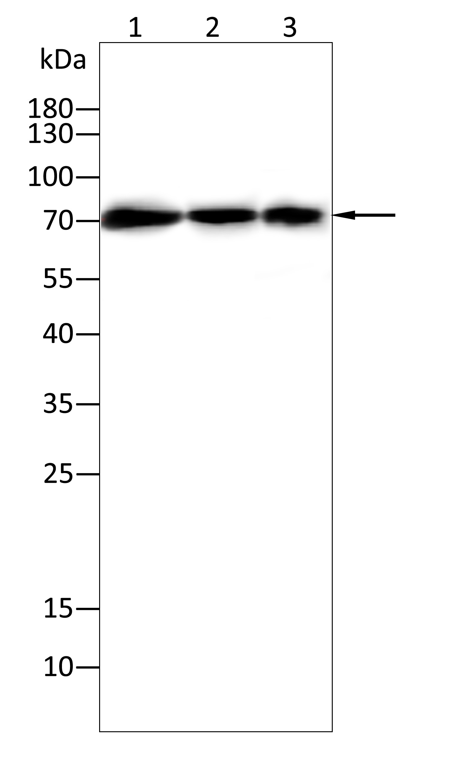
Western blot analysis using HSc70 mouse monoclonal antibody against Hepg2(1),A549(2),B16(3) cell lysate . 12% SDS-PAGE gel.Sample loading: 20μg /lane. Transfer the proteins onto a PVDF membrane (OM790003), and block it with TBST (OM750016) plus skimmed milk powder(OM750084) for one hour. Dilute the primary antibody with the antibody diluent (OM750012) at a ratio of 1:1000, and incubate it overnight at 4°C. Wash the membrane three times with TBST (OM750016), 5 minutes each time. At room temperature, dilute the secondary antibody, Goat Anti-Mouse IgG (H&L) - HRP(OM644366), at a ratio of 1:20000 and incubate for one hour. Wash the membrane three times with TBST (OM750016) again, 5 minutes each time. Use ECL (OM625701) for luminescence.staining time: 60SIHC
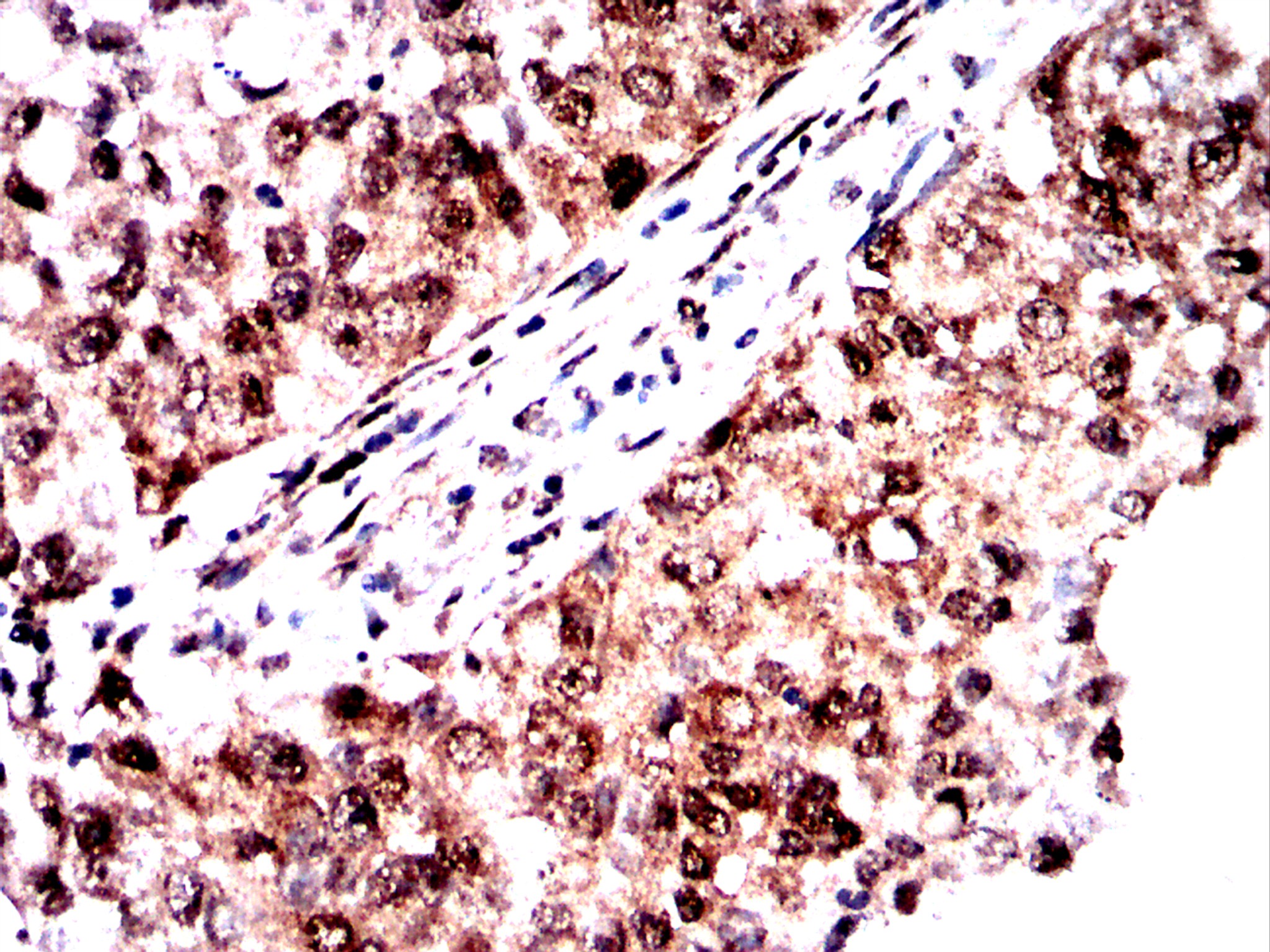
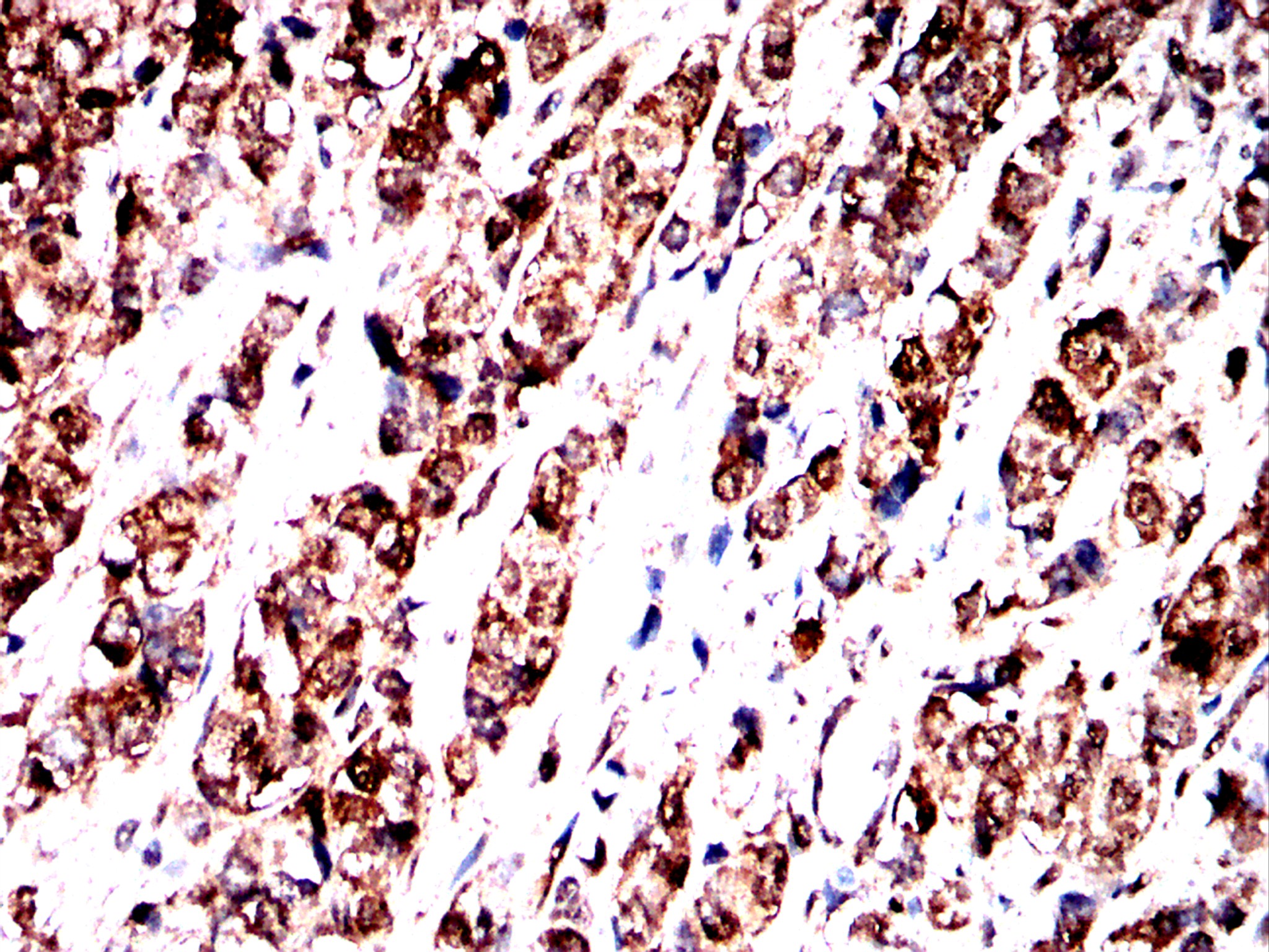
Immunohistochemical analysis of paraffin-embedded cervical cancer tissues using HSC70 antibody with DAB staining.Pre-treat the sections with heat-mediated antigen retrieval using sodium citrate buffer (pH 6.0) (OM750020) for 2 minutes. Wash the sections with ddH₂O and PBS (OM750003). Block the tissue with 10% non-immune goat serum(OM760028) at room temperature for 30 minutes. Incubate the tissue with the primary antibody diluted at a ratio of 1:1000 at 4°C overnight. At room temperature, dilute the secondary antibody, Goat Anti-Mouse IgG(H&L)-HRP (OM644366), at a ratio of 1:200 and incubate for one hour. Use DAB(OM760029)as the chromogenic agent. Counterstain the tissue with hematoxylin, and mount the tissue sections with neutral gum.IHC
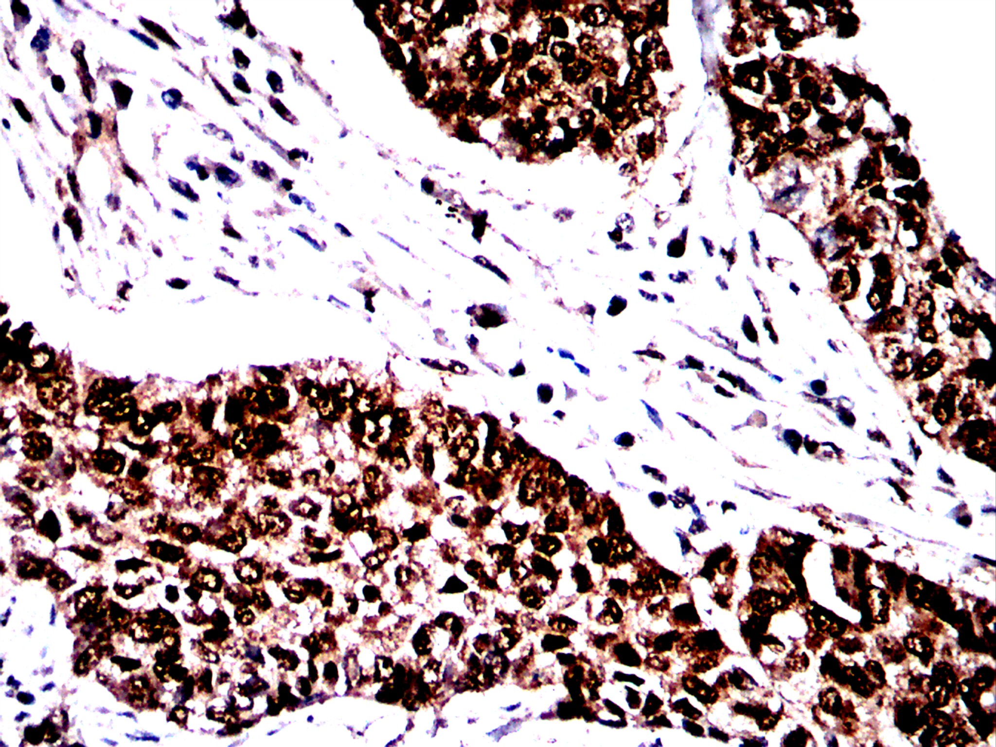
Immunohistochemical analysis of paraffin-embedded lung cancer tissues using HSC70 antibody with DAB staining.Pre-treat the sections with heat-mediated antigen retrieval using sodium citrate buffer (pH 6.0) (OM750020) for 2 minutes. Wash the sections with ddH₂O and PBS (OM750003). Block the tissue with 10% non-immune goat serum(OM760028) at room temperature for 30 minutes. Incubate the tissue with the primary antibody diluted at a ratio of 1:1000 at 4°C overnight. At room temperature, dilute the secondary antibody, Goat Anti-Mouse IgG(H&L)-HRP (OM644366), at a ratio of 1:200 and incubate for one hour. Use DAB(OM760029)as the chromogenic agent. Counterstain the tissue with hematoxylin, and mount the tissue sections with neutral gum.IHC
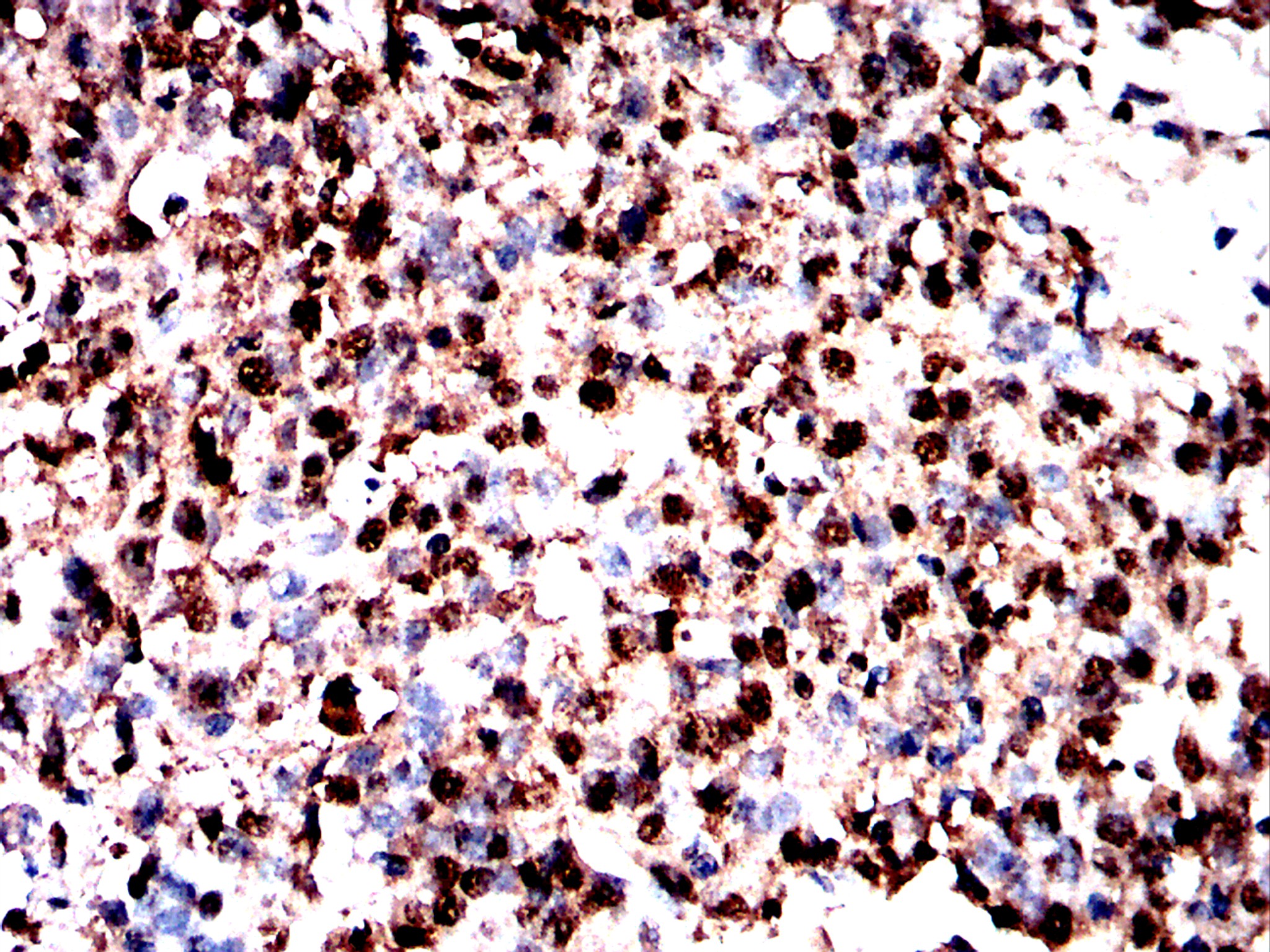
Immunohistochemical analysis of paraffin-embedded ovarian cancer tissues using HSC70 antibody with DAB staining.Pre-treat the sections with heat-mediated antigen retrieval using sodium citrate buffer (pH 6.0) (OM750020) for 2 minutes. Wash the sections with ddH₂O and PBS (OM750003). Block the tissue with 10% non-immune goat serum(OM760028) at room temperature for 30 minutes. Incubate the tissue with the primary antibody diluted at a ratio of 1:1000 at 4°C overnight. At room temperature, dilute the secondary antibody, Goat Anti-Mouse IgG(H&L)-HRP (OM644366), at a ratio of 1:200 and incubate for one hour. Use DAB(OM760029)as the chromogenic agent. Counterstain the tissue with hematoxylin, and mount the tissue sections with neutral gum.IHC
Immunohistochemical analysis of paraffin-embedded prostate cancer tissues using HSC70 antibody with DAB staining.Pre-treat the sections with heat-mediated antigen retrieval using sodium citrate buffer (pH 6.0) (OM750020) for 2 minutes. Wash the sections with ddH₂O and PBS (OM750003). Block the tissue with 10% non-immune goat serum(OM760028) at room temperature for 30 minutes. Incubate the tissue with the primary antibody diluted at a ratio of 1:1000 at 4°C overnight. At room temperature, dilute the secondary antibody, Goat Anti-Mouse IgG(H&L)-HRP (OM644366), at a ratio of 1:200 and incubate for one hour. Use DAB(OM760029)as the chromogenic agent. Counterstain the tissue with hematoxylin, and mount the tissue sections with neutral gum.IF-P
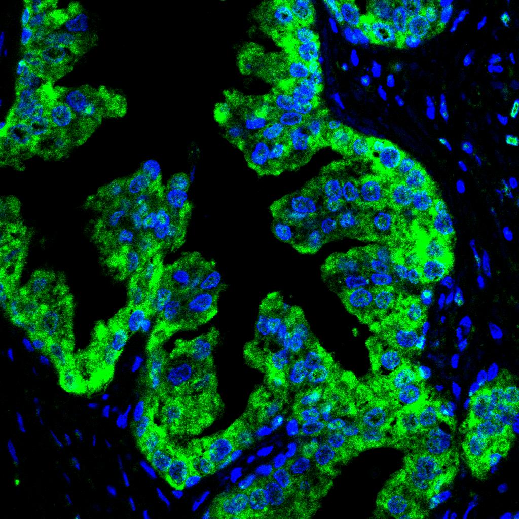
Immunohistochemical analysis of paraffin-embedded human prostate cancer tissues using HSC70 antibody with DAB staining.Pre-treat the sections with heat-mediated antigen retrieval using sodium citrate buffer (pH 6.0) (OM750020) for 2 minutes. Wash the sections with ddH₂O and PBS (OM750003). Block the tissue with 10% non-immune goat serum(OM760028) at room temperature for 30 minutes. Incubate the tissue with the primary antibody diluted at a ratio of 1:200 at 4°C overnight. At room temperature, dilute the secondary antibody, Goat Anti-Mouse IgG(H&L)-HRP (OM644366), at a ratio of 1:200 and incubate for one hour. Tyramine labeled with 488 (Green,OM642679) was used as chromogenic agent, and DAPI(Blue,OM642679) was used for double dyeing, and the anti-fluorescence attenuation tablets were sealed.ELISA
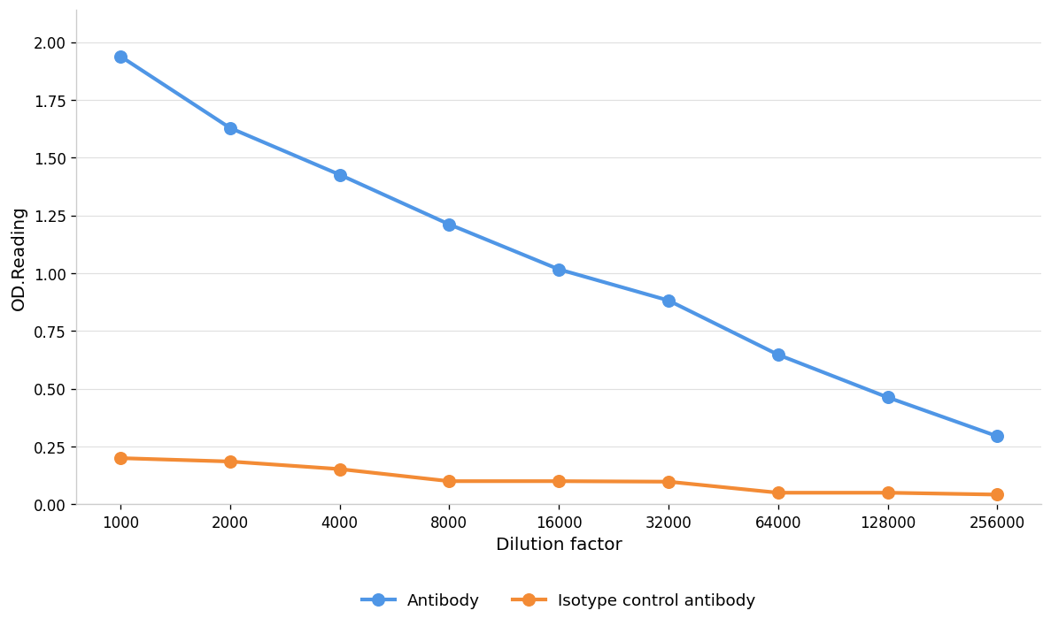
Blue line: Antibody; Orange line: Isotype control antibody| Product Name | Anti-HSc70 antibody |
|---|---|
| Antibody Type | Primary Antibodies |
| Immunogen | Recombinant protein |
| Clonality | Monoclonal |
|---|---|
| Isotype | IgG |
| Host Species | Mouse |
| Tested Applications | ELISAIF-PIHCWB |
| WB:1:1000 IHC:1:1000 IF-P:1:200-1:2000 ELISA:1:1000-1:10000 |
|
| Species Reactivity | HumanMouseRat |
| Concentration | 1mg/ml |
| Purification | Affinity purified |
| Gene Symbol | HSPA8 |
|---|---|
| Gene Synonyms | 2410008N15Rik 70kDa APG-2 HEL-33 HEL-S-5a HEL-S-72p HS24/P52 HSC54 HSC70 hsc70.I HSC71 Hsc73 Hsp110 HSP70 Hsp70RY HSP71 HSP73 HSPA1 HSPA10 hspa1l HSPH2 I79_004332 I79_022974 irp94 LAP-1 LAP1 mKIAA4025 NIP71 RY XELAEV_18037286mg xhsp70 |
| Gene Full Name | heat shock protein family A (Hsp70) member 8 |
| Alternative Names | 70 kDa heat-shock-like protein constitutive heat shock protein 70 epididymis luminal protein 33 epididymis secretory sperm binding protein Li 5a epididymis secretory sperm binding protein Li 72p Heat shock 70 kDa protein 4 Heat shock 70 kDa protein 8 heat shock 70-related protein APG-2 heat shock 70kd protein 10 heat shock 70kd protein 10 (HSC71) heat shock 70kD protein 4 heat shock 70kD protein 8 heat shock 70kDa protein 1-like heat shock 70kDa protein 4 heat shock 70kDa protein 8 heat shock cognate 70 heat shock cognate 71 kD protein heat shock cognate hsc73 heat shock cognate protein 54 Heat shock cognate protein 70 heat shock protein 4 heat shock protein 70 cognate heat shock protein 8 heat shock protein A8 heat shock protein Apg-2 heat shock protein cognate 70 heat shock protein family A member 4 heat shock protein family A member 8 heat shock protein family H member 2 heat shock protein 110 kDa Hsc70 ATPase hsp70 RY HSPA4/HSPA8 IRP94 ischemia responsive 94 kDa protein lipopolysaccharide-associated protein 1 LPS-associated protein 1 N-myristoyltransferase inhibitor protein 71 |
| Molecular Weight(MW) | 70kDa |
| Function | The HSP 70 family is composed of four highly conserved proteins: HSP 70, HSC 70, GRP 75 and GRP 78. These proteins serve a variety of roles: they act as molecular chaperones facilitating the assembly of multi-protein complexes, participate in the translocation of polypeptides across cell membranes and to the nucleus, and aid in the proper folding of nascent polypeptide chains. All members of the family, except HSP 70, are constitutively expressed in primate cells. HSP 70 expression is strongly induced in response to heat stress. HSP 70 and HSC 70 play key roles in the cytosolic endoplasmic reticulum and mitochondrial import machinery, and are found in both the cytosol and nucleus of mammalian cells. Both HSP 70 and HSC 70 are involved in the chaperoning of nascent polypeptide chains and in protecting cells against the accumulation of improperly folded proteins. GRP 78 is localized in the endoplasmic reticulum, where it receives imported secretory proteins and is involved in the folding and translocation of nascent peptide chains. GRP 75 expression is restricted to the mitochondrial matrix and aids in the translocation and folding of nascent polypeptide chains of both nuclear and mitochondrial origin. GRP 75 and GRP 78 are unresponsive to heat stress and are induced by glucose deprivation. It has been postulated that members of the HSP 70 family act as force-generating motors, relying on the hydrolysis of ATP for their activity. |
| Cellular Localization | Cytoplasm |
| SwissProt ID | P11142 |
|---|
WB
Western blot analysis using HSc70 mouse monoclonal antibody against Hepg2(1),A549(2),B16(3) cell lysate . 12% SDS-PAGE gel.Sample loading: 20μg /lane. Transfer the proteins onto a PVDF membrane (OM790003), and block it with TBST (OM750016) plus skimmed milk powder(OM750084) for one hour. Dilute the primary antibody with the antibody diluent (OM750012) at a ratio of 1:1000, and incubate it overnight at 4°C. Wash the membrane three times with TBST (OM750016), 5 minutes each time. At room temperature, dilute the secondary antibody, Goat Anti-Mouse IgG (H&L) - HRP(OM644366), at a ratio of 1:20000 and incubate for one hour. Wash the membrane three times with TBST (OM750016) again, 5 minutes each time. Use ECL (OM625701) for luminescence.staining time: 60S
IHC
Immunohistochemical analysis of paraffin-embedded cervical cancer tissues using HSC70 antibody with DAB staining.Pre-treat the sections with heat-mediated antigen retrieval using sodium citrate buffer (pH 6.0) (OM750020) for 2 minutes. Wash the sections with ddH₂O and PBS (OM750003). Block the tissue with 10% non-immune goat serum(OM760028) at room temperature for 30 minutes. Incubate the tissue with the primary antibody diluted at a ratio of 1:1000 at 4°C overnight. At room temperature, dilute the secondary antibody, Goat Anti-Mouse IgG(H&L)-HRP (OM644366), at a ratio of 1:200 and incubate for one hour. Use DAB(OM760029)as the chromogenic agent. Counterstain the tissue with hematoxylin, and mount the tissue sections with neutral gum.
IHC
Immunohistochemical analysis of paraffin-embedded lung cancer tissues using HSC70 antibody with DAB staining.Pre-treat the sections with heat-mediated antigen retrieval using sodium citrate buffer (pH 6.0) (OM750020) for 2 minutes. Wash the sections with ddH₂O and PBS (OM750003). Block the tissue with 10% non-immune goat serum(OM760028) at room temperature for 30 minutes. Incubate the tissue with the primary antibody diluted at a ratio of 1:1000 at 4°C overnight. At room temperature, dilute the secondary antibody, Goat Anti-Mouse IgG(H&L)-HRP (OM644366), at a ratio of 1:200 and incubate for one hour. Use DAB(OM760029)as the chromogenic agent. Counterstain the tissue with hematoxylin, and mount the tissue sections with neutral gum.
IHC
Immunohistochemical analysis of paraffin-embedded ovarian cancer tissues using HSC70 antibody with DAB staining.Pre-treat the sections with heat-mediated antigen retrieval using sodium citrate buffer (pH 6.0) (OM750020) for 2 minutes. Wash the sections with ddH₂O and PBS (OM750003). Block the tissue with 10% non-immune goat serum(OM760028) at room temperature for 30 minutes. Incubate the tissue with the primary antibody diluted at a ratio of 1:1000 at 4°C overnight. At room temperature, dilute the secondary antibody, Goat Anti-Mouse IgG(H&L)-HRP (OM644366), at a ratio of 1:200 and incubate for one hour. Use DAB(OM760029)as the chromogenic agent. Counterstain the tissue with hematoxylin, and mount the tissue sections with neutral gum.
IHC
Immunohistochemical analysis of paraffin-embedded prostate cancer tissues using HSC70 antibody with DAB staining.Pre-treat the sections with heat-mediated antigen retrieval using sodium citrate buffer (pH 6.0) (OM750020) for 2 minutes. Wash the sections with ddH₂O and PBS (OM750003). Block the tissue with 10% non-immune goat serum(OM760028) at room temperature for 30 minutes. Incubate the tissue with the primary antibody diluted at a ratio of 1:1000 at 4°C overnight. At room temperature, dilute the secondary antibody, Goat Anti-Mouse IgG(H&L)-HRP (OM644366), at a ratio of 1:200 and incubate for one hour. Use DAB(OM760029)as the chromogenic agent. Counterstain the tissue with hematoxylin, and mount the tissue sections with neutral gum.
IF-P
Immunohistochemical analysis of paraffin-embedded human prostate cancer tissues using HSC70 antibody with DAB staining.Pre-treat the sections with heat-mediated antigen retrieval using sodium citrate buffer (pH 6.0) (OM750020) for 2 minutes. Wash the sections with ddH₂O and PBS (OM750003). Block the tissue with 10% non-immune goat serum(OM760028) at room temperature for 30 minutes. Incubate the tissue with the primary antibody diluted at a ratio of 1:200 at 4°C overnight. At room temperature, dilute the secondary antibody, Goat Anti-Mouse IgG(H&L)-HRP (OM644366), at a ratio of 1:200 and incubate for one hour. Tyramine labeled with 488 (Green,OM642679) was used as chromogenic agent, and DAPI(Blue,OM642679) was used for double dyeing, and the anti-fluorescence attenuation tablets were sealed.
ELISA
Blue line: Antibody; Orange line: Isotype control antibody| Application Notes | WB:1:1000 IHC:1:1000 IF-P:1:200-1:2000 ELISA:1:1000-1:10000 |
|---|
| Form | Liquid |
|---|---|
| Storage Instructions | Shipped at 4°C. Store at +4°C short term (1-2 weeks). Store at -20°C long term. Avoid freeze / thaw cycle. |
| Storage Buffer | Purified antibody in PBS with 0.05% sodium azide,40% Glycerol. |


