WB
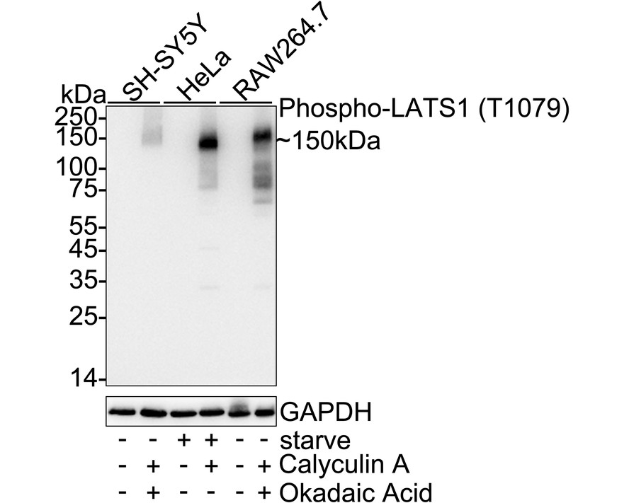
Western blot analysis of Phospho-LATS1 (T1079) on different lysates with Rabbit anti-Phospho-LATS1 (T1079) antibody at 1/2,000 dilution. Lane 1: SH-SY5Y cell lysate, Lane 2: SH-SY5Y treated with 200nM Calyculin A and 1μM Okadaic Acid for 1 hour cell lysate, Lane 3: HeLa starved for 3 hours cell lysate, Lane 4: HeLa starved for 3 hours then treated with 100nM Calyculin A for 30 minutes cell lysate, Lane 5: RAW264.7 cell lysate, Lane 6: RAW264.7 treated with 200nM Calyculin A and 1μM Okadaic Acid for 1 hour cell lysate, Lysates/proteins at 20 µg/Lane. Exposure time: 2 minutes; 4-20% SDS-PAGE gel. Proteins were transferred to a PVDF membrane and blocked with 5% NFDM/TBST for 1 hour at room temperature. The primary antibody at 1/2,000 dilution was used in 5% NFDM/TBST at 4℃ overnight. Goat Anti-Rabbit IgG - HRP Secondary Antibodyat 1/50,000 dilution was used for 1 hour at room temperature.IP
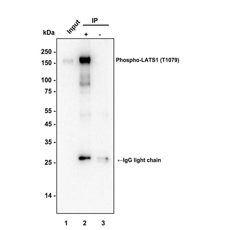
Phospho-LATS1 (T1079) was immunoprecipitated from 0.2 mg HeLa treated with 50mM Calyculin A for 3 hours cell lysate with Rabbit anti-Phospho-LATS1 (T1079) antibody at 2 µg/10 µl beads. Western blot was performed from the immunoprecipitate using Rabbit anti-Phospho-LATS1 (T1079) antibody at 1/2,000 dilution. HRP Conjugated Anti-Rabbit IgG for IP Nano-secondary antibody at 1/5,000 dilution was used for 1 hour at room temperature. Lane 1: HeLa treated with 50mM Calyculin A for 3 hours cell lysate (input), Lane 2: Rabbit anti-Phospho-LATS1 (T1079) antibody IP in HeLa treated with 50mM Calyculin A for 3 hours cell lysate, Lane 3: Rabbit IgG instead of Rabbit anti-Phospho-LATS1 (T1079) antibody in HeLa treated with 50mM Calyculin A for 3 hours cell lysate. Blocking/Dilution buffer: 5% NFDM/TBST Exposure time: 1 minute 34 seconds.| Product Name | Phospho-LATS1 (T1079) Recombinant Rabbit Monoclonal Antibody |
|---|---|
| Antibody Type | Primary Antibodies |
| Immunogen | Synthetic phospho-peptide corresponding to residues surrounding Thr1079 of Human LATS1. |
| Modification | p-T1079 |
| Clonality | Monoclonal |
|---|---|
| Isotype | IgG |
| Host Species | Rabbit |
| Tested Applications | IPWB |
| WB:1:2000 IP:1-2μg/sample |
|
| Species Reactivity | HumanMouse |
| Concentration | 1mg/ml |
| Purification | Protein A |
| Gene Symbol | LATS1 |
|---|---|
| Gene Synonyms | wts WARTS |
| Gene Full Name | large tumor suppressor kinase 1 |
| Gene Summary | The protein encoded by this gene is a putative serine/threonine kinase that localizes to the mitotic apparatus and complexes with cell cycle controller CDC2 kinase in early mitosis. The protein is phosphorylated in a cell-cycle dependent manner, with late prophase phosphorylation remaining through metaphase. The N-terminal region of the protein binds CDC2 to form a complex showing reduced H1 histone kinase activity, indicating a role as a negative regulator of CDC2/cyclin A. In addition, the C-terminal kinase domain binds to its own N-terminal region, suggesting potential negative regulation through interference with complex formation via intramolecular binding. Biochemical and genetic data suggest a role as a tumor suppressor. This is supported by studies in knockout mice showing development of soft-tissue sarcomas, ovarian stromal cell tumors and a high sensitivity to carcinogenic treatments. [provided by RefSeq, Apr 2017] |
| Molecular Weight(MW) | 127 kDa(Observed band size: 150 kDa) |
| Cellular Localization | Cytoplasm, cytoskeleton, microtubule organizing center, centrosome, spindle, microtubule organizing center, spindle pole body. |
WB
Western blot analysis of Phospho-LATS1 (T1079) on different lysates with Rabbit anti-Phospho-LATS1 (T1079) antibody at 1/2,000 dilution. Lane 1: SH-SY5Y cell lysate, Lane 2: SH-SY5Y treated with 200nM Calyculin A and 1μM Okadaic Acid for 1 hour cell lysate, Lane 3: HeLa starved for 3 hours cell lysate, Lane 4: HeLa starved for 3 hours then treated with 100nM Calyculin A for 30 minutes cell lysate, Lane 5: RAW264.7 cell lysate, Lane 6: RAW264.7 treated with 200nM Calyculin A and 1μM Okadaic Acid for 1 hour cell lysate, Lysates/proteins at 20 µg/Lane. Exposure time: 2 minutes; 4-20% SDS-PAGE gel. Proteins were transferred to a PVDF membrane and blocked with 5% NFDM/TBST for 1 hour at room temperature. The primary antibody at 1/2,000 dilution was used in 5% NFDM/TBST at 4℃ overnight. Goat Anti-Rabbit IgG - HRP Secondary Antibodyat 1/50,000 dilution was used for 1 hour at room temperature.
IP
Phospho-LATS1 (T1079) was immunoprecipitated from 0.2 mg HeLa treated with 50mM Calyculin A for 3 hours cell lysate with Rabbit anti-Phospho-LATS1 (T1079) antibody at 2 µg/10 µl beads. Western blot was performed from the immunoprecipitate using Rabbit anti-Phospho-LATS1 (T1079) antibody at 1/2,000 dilution. HRP Conjugated Anti-Rabbit IgG for IP Nano-secondary antibody at 1/5,000 dilution was used for 1 hour at room temperature. Lane 1: HeLa treated with 50mM Calyculin A for 3 hours cell lysate (input), Lane 2: Rabbit anti-Phospho-LATS1 (T1079) antibody IP in HeLa treated with 50mM Calyculin A for 3 hours cell lysate, Lane 3: Rabbit IgG instead of Rabbit anti-Phospho-LATS1 (T1079) antibody in HeLa treated with 50mM Calyculin A for 3 hours cell lysate. Blocking/Dilution buffer: 5% NFDM/TBST Exposure time: 1 minute 34 seconds.| Application Notes | WB:1:2000 IP:1-2μg/sample |
|---|
| Form | Liquid |
|---|---|
| Storage Instructions | Shipped at 4℃. Store at +4℃ short term (1-2 weeks). It is recommended to aliquot into single-use upon delivery. Store at -20℃ long term. |
| Storage Buffer | PBS (pH7.4), 0.1% BSA, 40% Glycerol. Preservative: 0.05% Sodium Azide. |


