WB
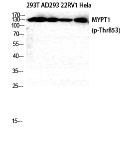
Western Blot analysis of 293T AD293 22RV1 HELA cells using Phospho-MYPT1 (T853) Polyclonal Antibody diluted at 1:2000.IHC
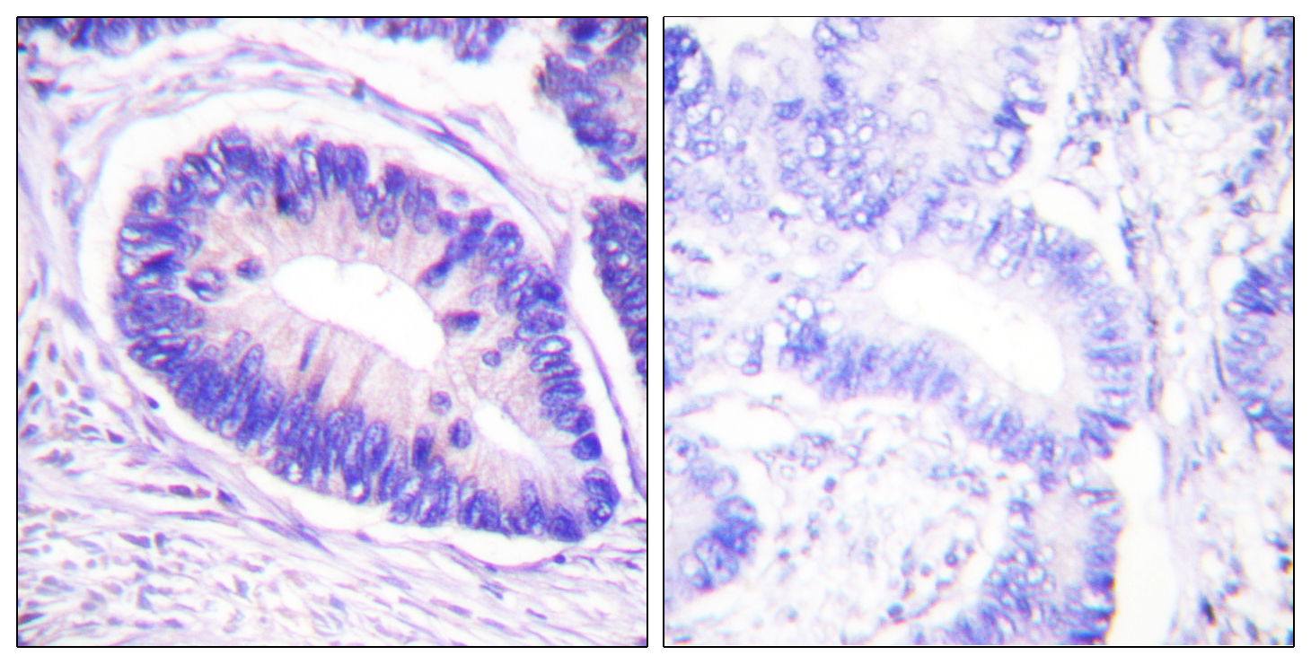
Immunohistochemistry analysis of paraffin-embedded human colon carcinoma, using MYPT1 (Phospho-Thr853) Antibody. The picture on the right is blocked with the phospho peptide.ICC/IF
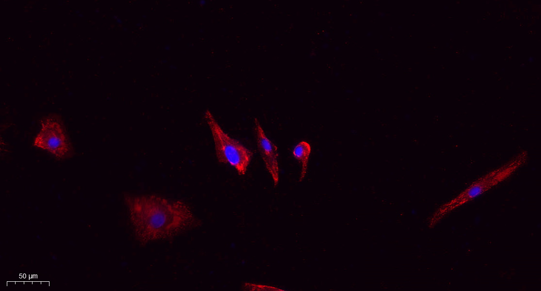
Immunofluorescence analysis of A549. 1,primary Antibody(red) was diluted at 1:200(4℃ overnight). 2, Goat Anti Rabbit IgG (H&L) - Alexa Fluor 594 Secondary antibody was diluted at 1:1000(room temperature, 50min).3, DAPI(blue) 10min.ICC/IF
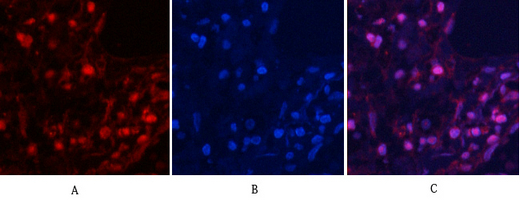
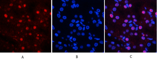
Immunofluorescence analysis of human-lung tissue. 1,MYPT1 (phospho Thr853) Polyclonal Antibody(red) was diluted at 1:200(4℃,overnight). 2, Cy3 labled Secondary antibody was diluted at 1:300(room temperature, 50min).3, Picture B: DAPI(blue) 10min. Picture A:Target. Picture B: DAPI. Picture C: merge of A+B.ICC/IF
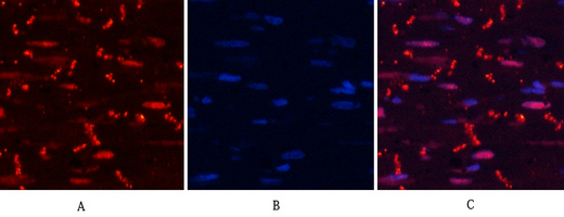
Immunofluorescence analysis of rat-heart tissue. 1,MYPT1 (phospho Thr853) Polyclonal Antibody(red) was diluted at 1:200(4℃,overnight). 2, Cy3 labled Secondary antibody was diluted at 1:300(room temperature, 50min).3, Picture B: DAPI(blue) 10min. Picture A:Target. Picture B: DAPI. Picture C: merge of A+B.ICC/IF
Immunofluorescence analysis of rat-kidney tissue. 1,MYPT1 (phospho Thr853) Polyclonal Antibody(red) was diluted at 1:200(4℃,overnight). 2, Cy3 labled Secondary antibody was diluted at 1:300(room temperature, 50min).3, Picture B: DAPI(blue) 10min. Picture A:Target. Picture B: DAPI. Picture C: merge of A+B.| Product Name | MYPT1 Rabbit pAb |
|---|---|
| Antibody Type | Primary Antibodies |
| Immunogen | The antiserum was produced against synthesized peptide derived from human MYPT1 around the phosphorylation site of Thr853. AA range:621-670 |
| Clonality | Polyclonal |
|---|---|
| Isotype | IgG |
| Host Species | Rabbit |
| Tested Applications | ICC/IFIHCWB |
| WB:1:500-1:2000 IHC:1:100-1:300 ICC/IF:1:50-1:200 |
|
| Species Reactivity | HumanMouseRat |
| Concentration | 1mg/ml |
| Purification | Protein A |
| Gene Symbol | PPP1R12A |
|---|---|
| Gene Synonyms | MBS GUBS M130 MYPT1 |
| Gene Full Name | protein phosphatase 1 regulatory subunit 12A |
| Gene Summary | Myosin phosphatase target subunit 1, which is also called the myosin-binding subunit of myosin phosphatase, is one of the subunits of myosin phosphatase. Myosin phosphatase regulates the interaction of actin and myosin downstream of the guanosine triphosphatase Rho. The small guanosine triphosphatase Rho is implicated in myosin light chain (MLC) phosphorylation, which results in contraction of smooth muscle and interaction of actin and myosin in nonmuscle cells. The guanosine triphosphate (GTP)-bound, active form of RhoA (GTP.RhoA) specifically interacted with the myosin-binding subunit (MBS) of myosin phosphatase, which regulates the extent of phosphorylation of MLC. Rho-associated kinase (Rho-kinase), which is activated by GTP. RhoA, phosphorylated MBS and consequently inactivated myosin phosphatase. Overexpression of RhoA or activated RhoA in NIH 3T3 cells increased phosphorylation of MBS and MLC. Thus, Rho appears to inhibit myosin phosphatase through the action of Rho-kinase. Several transcript variants encoding different isoforms have been found for this gene. [provided by RefSeq, Jan 2009] |
| Alternative Names | PPP1R12A MBS MYPT1 Protein phosphatase 1 regulatory subunit 12A Myosin phosphatase-targeting subunit 1 Myosin phosphatase target subunit 1 Protein phosphatase myosin-binding subunit |
| Molecular Weight(MW) | 130 kDa |
| Cellular Localization | Cytoplasm. |
WB
Western Blot analysis of 293T AD293 22RV1 HELA cells using Phospho-MYPT1 (T853) Polyclonal Antibody diluted at 1:2000.
IHC
Immunohistochemistry analysis of paraffin-embedded human colon carcinoma, using MYPT1 (Phospho-Thr853) Antibody. The picture on the right is blocked with the phospho peptide.
ICC/IF
Immunofluorescence analysis of A549. 1,primary Antibody(red) was diluted at 1:200(4℃ overnight). 2, Goat Anti Rabbit IgG (H&L) - Alexa Fluor 594 Secondary antibody was diluted at 1:1000(room temperature, 50min).3, DAPI(blue) 10min.
ICC/IF
Immunofluorescence analysis of human-lung tissue. 1,MYPT1 (phospho Thr853) Polyclonal Antibody(red) was diluted at 1:200(4℃,overnight). 2, Cy3 labled Secondary antibody was diluted at 1:300(room temperature, 50min).3, Picture B: DAPI(blue) 10min. Picture A:Target. Picture B: DAPI. Picture C: merge of A+B.
ICC/IF
Immunofluorescence analysis of rat-heart tissue. 1,MYPT1 (phospho Thr853) Polyclonal Antibody(red) was diluted at 1:200(4℃,overnight). 2, Cy3 labled Secondary antibody was diluted at 1:300(room temperature, 50min).3, Picture B: DAPI(blue) 10min. Picture A:Target. Picture B: DAPI. Picture C: merge of A+B.
ICC/IF
Immunofluorescence analysis of rat-kidney tissue. 1,MYPT1 (phospho Thr853) Polyclonal Antibody(red) was diluted at 1:200(4℃,overnight). 2, Cy3 labled Secondary antibody was diluted at 1:300(room temperature, 50min).3, Picture B: DAPI(blue) 10min. Picture A:Target. Picture B: DAPI. Picture C: merge of A+B.| Application Notes | WB:1:500-1:2000 IHC:1:100-1:300 ICC/IF:1:50-1:200 |
|---|
| Form | Liquid |
|---|---|
| Storage Instructions | -15°C to -25°C/1 year(Do not lower than -25°C) |
| Storage Buffer | PBS, 50% glycerol, 0.05% Proclin 300, 0.05%BSA |


